 Loading...
Do you have a question about the Welch Allyn 11720 and is the answer not in the manual?
| Model Number | 11720 |
|---|---|
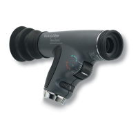
| Category | Ophthalmoscope |
| Illumination | Halogen |
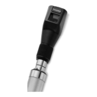
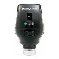
| Head Type | Standard |
| Apertures | Small, Large, Fixation |
| Filters | Red-Free |
Details various apertures like micro spot, large spot, fixation, slit, and cobalt blue filters for examination.
Explains red-free and crossed linear polarizing filters, their functions, and how to engage them.
Describes using the ophthalmoscope for examining other ocular structures like the cornea and iris.
Illustrates the eye's structure and explains how ophthalmoscope lenses aid clear vision for different refractive errors.
Provides a step-by-step guide for performing a fundus examination, including patient positioning and observation techniques.
Offers methods, including filter use and light beam direction, to minimize corneal reflection for clearer retinal views.
Describes the typical appearance of the optic disc, retina, and blood vessels in a healthy eye.
Details advanced malignant hypertensive retinopathy, noting disc edema, flame hemorrhages, and attenuated arterioles.
Explains the presence of new vessels on the disc surface, hemorrhages, and dilated veins in this condition.
Describes an elevated, edematous optic disc with blurred margins and engorged, tortuous vessels.
Covers the appearance of a benign choroidal nevus, including its slate gray color, overlying drusen, and normal vessels.
Details gray elevation with folds, tortuous and elevated vessels, and the overall appearance of detached retina.