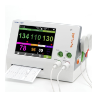
10
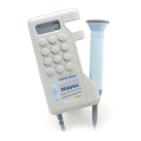
Operation
3. Operation
Refer to diagram on page 3 for Doppler Measuring sites and
Recommended Probes.
To connect the probe, align the arrow on the connector with the slot on the
probe and push fi rmly.
To disconnect the probe, pull the connector sharply. DO NOT pull the cable.
Note: During use, an automatic noise reduction feature operates on low
level signals to improve sound quality.
Coupling Gel
Use water based ultrasound gel ONLY.
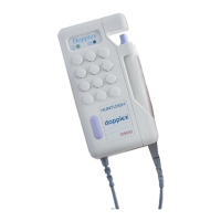
3.1 Vascular Mode
The Multi Dopplex II/Super Dopplex II Dopplers (MD2/SD2) will select vascular
mode when a vascular probe is connected to the control unit.
Vascular Probes
Five probes are available for vascular examinations:
VP4HS
4MHz ±1% for deep lying vessels
VP5HS
5MHz ±1% for deep lying vessels and oedematous limbs
VP8HS
8MHz ±1% for peripheral vessels
VP10HS
10MHz ±1% for specialist superfi cial applications.
EZ8
8MHZ ±1% “Widebeam” for peripheral vessels.
In this mode, bi-directional blood fl ow rate and direction are indicated on bar
graphs (4 levels in each direction) and blood fl ow is audible in the loudspeaker.
Probe frequency is displayed together with the bar graphs.
Clinical Use
Apply a liberal amount of gel on the site to be examined. Place the probe at
45° to the skin surface over the vessel to be examined. Adjust the position of
the probe to obtain the loudest audio signal. High pitched pulsatile sounds are
emitted from arteries while veins emit a non-pulsatile sound similar to a rushing
wind.
For best results, keep the probe as still as possible once the optimum position
has been found. Adjust the audio volume as required.
 Loading...