4
The
PanOptic
™ Ophthalmoscope
Transparency of the cornea, lens and vitreous humor permits the
practitioner to directly view arteries, veins, and the optic nerve of
the retina.
Direct observation of the structures of the fundus through a PanOp-
tic Ophthalmoscope may show disease of the eye itself or may
reveal abnormalities indicative of disease elsewhere in the body.
Among the most important of these are vascular changes due to dia-
betes or hypertension and swelling of the optic nerve head due to
papilledema or optic neuritis. In this sense, the eye serves as a win-
dow through which many valuable clinical evaluations may be
made.
When a preliminary diagnosis of an imminently dangerous eye con-
dition, such as acute (angle-closure) glaucoma or retinal detach-
ment, is made by the examiner, prompt referral to an eye specialist
may prevent irreversible damage. Or, when distressing but less
urgent conditions, such as visual impairment due to cataract or vit-
reous floaters, are recognized, the patient can be reassured and
referred.
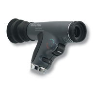
A
B
C
D
E
F
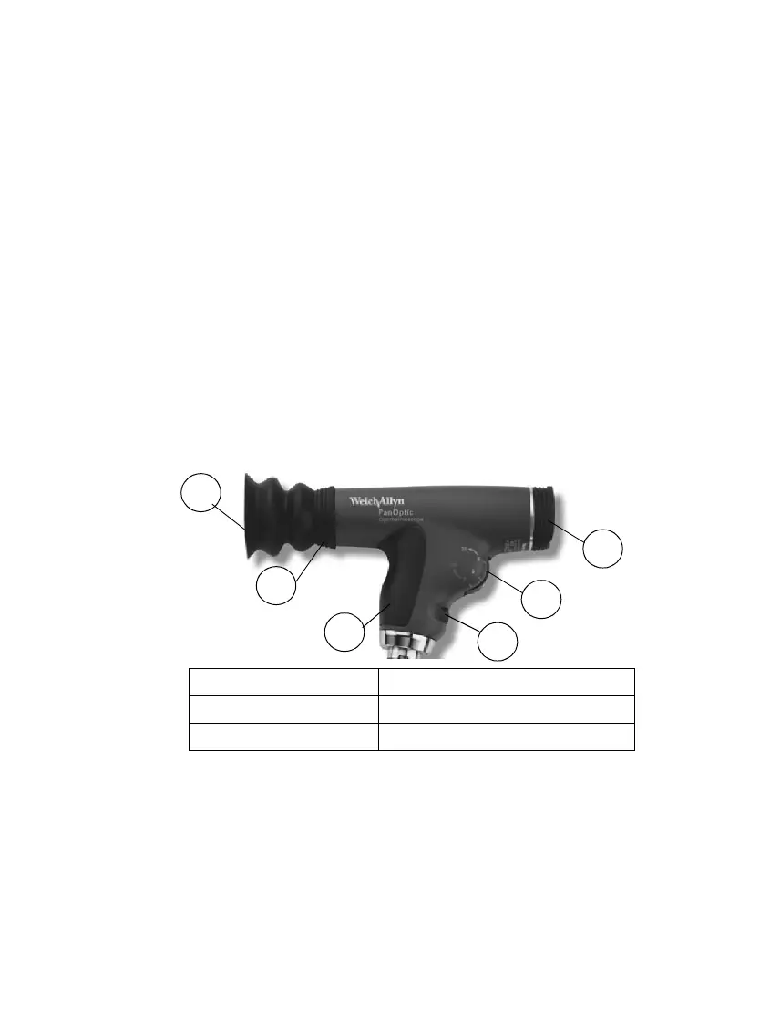
A Patient Eyecup D Aperture/Filter Dial
B Patient’s Side E Focusing Wheel
C Soft Grip Handle F Practitioner’s Side Brow Rest
 Loading...