70
Chapter 4 Operation
EVIS EXERA II GIF/CF/PCF TYPE 180 Series OPERATION MANUAL
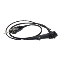
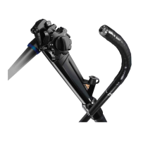
4.2 Using endo-therapy accessories
For more information on combining the endoscope with particular endo-therapy
accessories, refer to the “System chart” in the Appendix and the instruction
manuals of the accessories. Refer to the accessories’ instruction manuals for
operating instructions.
• Do not use the GIF-N180 for high-frequency cauterization or
laser cauterization treatment. Otherwise, patient injury or
equipment damage may result.
• When using endo-therapy accessories, keep the distance
between the distal end of the endoscope and the mucous
membrane greater than the endoscope’s minimum visible
distance so that the endo-therapy accessory remains visible
in the endoscopic image. If the distal end of the endoscope is
placed closer than its own minimum visible distance, the
position of the accessory cannot be seen in the endoscopic
image, which could cause serious patient injury and/or
equipment damage. The minimum visible distance depends
on the type of endoscope being used. Refer to Section 2.3,
“Specifications” on page 24.
• When inserting or withdrawing an endo-therapy accessory,
confirm that its distal end is closed or completely retracted
into the sheath. Slowly insert or withdraw the endo-therapy
accessory straight into/from the slit of the biopsy valve.
Otherwise, the biopsy valve may be damaged and pieces of
it could fall off.
• If the insertion or withdrawal of endo-therapy accessories is
difficult, straighten the bending section as much as possible
without losing the endoscopic image. Inserting or
withdrawing endo-therapy accessories with excessive force
may damage the instrument channel or endo-therapy
accessories cause some parts to fall off and/or cause patient
injury.
• If the distal end of an endo-therapy accessory is not visible in
the endoscopic image, do not open the distal end or extend
the needle of the instrument. This could cause patient injury,
bleeding, perforation and/or equipment damage.
 Loading...