Image Optimization 5-67
5.13.1 Basic Procedures for Contrast Imaging
To perform a successful contrast imaging, you should start with an optimized 2D image
and have the target region in mind. To perform a contrast imaging:
1. Select an appropriate probe, and perform 2D imaging to obtain the target image, and
then fix the probe.
2. Tap [Contrast] to enter the contrast imaging mode.
3. Adjust the acoustic power experientially to obtain a good image.
Tap [Dual Live] to be “On” to activate the dual live function. Observe the tissue image
to find the target view.
4. Inject the contrast agent, and set [Timer 1] at “ON” to start the contrast timing. When
the timer begins to work, the time will be displayed on the screen.
5. Observe the image, use the touch screen button of [Pro Capture] and [Retro Capture]
or the user-defined key to save the images. Press <Freeze> to end the live capture.
Perform several live captures if there are more than one interested sections.
6. At the end of a contrast imaging, set [Timer 1] as “OFF” to exit the timing function.
Perform procedures 3-5 if necessary.
For every single contrast imaging procedure, use [Timer 2] for timing.
If necessary, activate destruction function by tapping [Destruct] as “ON” to destruct
the micro-bubbles left by the last contrast imaging; or to observe the reinfusion effect
in a continuous agent injecting process.
7. Exit contrast imaging.
Press <B> button to return to B mode.
5.13.1.1 Parameter Area Display
When entering contrast imaging mode, the screen displays the contrast image, and if
[Dual Live] item on the touch screen is “ON”, both the contrast image (marked with “
”)
and tissue image (marked with “ ”) are displayed (the two window position can be
changed). Parameter area displays as follows:
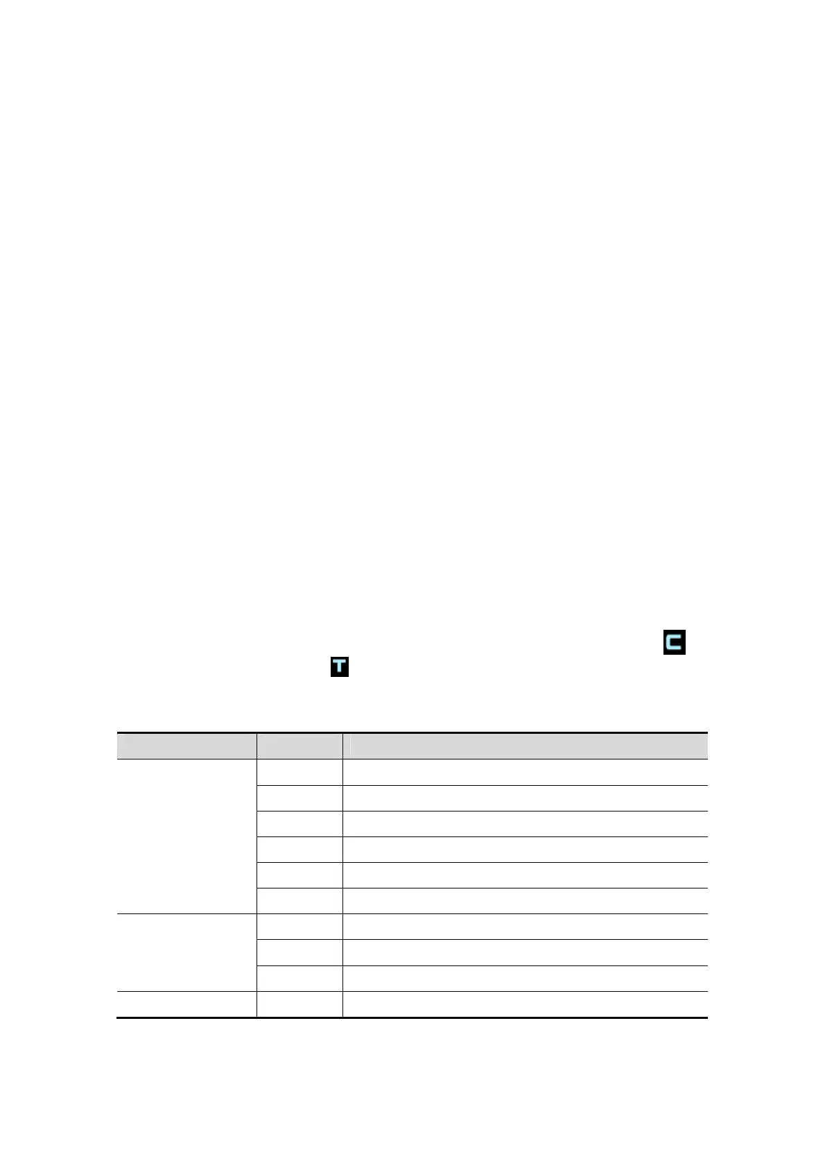
Type Parameter Description
Contrast
FC Contrast frequency.
D Depth.
G Gain.
FR Frame rate.
DR Dynamic Range.
iTouch iTouch status
Tissue
G Gain
DR Dynamic Range.
iTouch iTouch status
Zoom Z Magnification factor

 Loading...