16 - 4 Operator’s Manual
16 Probes and Biopsy
Abdominal use of C5-1s and SC5-1Ns includes gynecology application region.
16.1.1 Probe Functions by Part
The basic structures and corresponding functions of probes are basically the same; take the
following probe as an example.
SP5-1Ns Body surface Abdominal,
Pediatric,
Neonatal
Cephalic, Adult
Cephalic,
Thoracic/Pleural,
Cardiac Adult,
Cardiac Pediatric
B, M, PW, CW, Color,
Power, Combined
modes (B+M, PW+B,
Color + B, Power +
B, PW +Color+ B,
Power + PW +B),
Tissue Harmonic
Imaging, iScape, TDI,
Color M, Biopsy
Guidance, Smart 3D,
Contrast imaging
(Contrast agent for
LVO)
Table 16-1 Available probes
Probe Model Region Applied Intended Use Imaging Mode Probe Figure
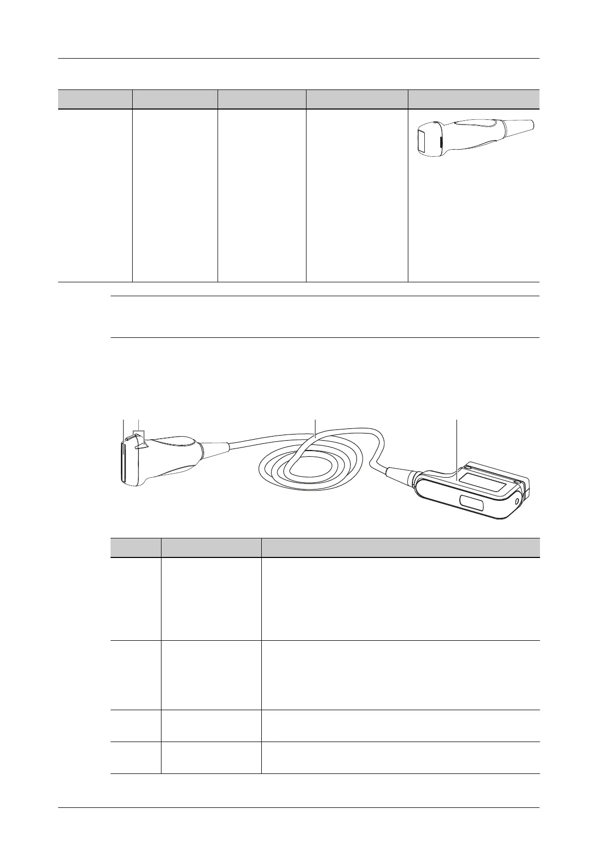
No. Item Description
1. Probe head Converts the electrical signal into an ultrasonic signal,
focusing the sound beams in a given direction; meanwhile, it
receives the reflected ultrasonic signal and converts it into an
electrical signal for transmission over the cable. The lens on
the surface is the acoustic lens. Apply ultrasound gel on the
acoustic lens for correct operation.
2. Needle-guided
bracket fix tabs and
grooves
Provides mounting support of the needle-guided bracket.
NOTE:
This structure of probes in the figure above may vary with
the matched needle-guided brackets.
3. Probe cable Transmits electrical signals between the probe body and
connector.
4. Probe connector Connects the probe and cable to the ultrasonic diagnostic
system.
 Loading...