16 Probes and Biopsy
Operator’s Manual 16 - 5
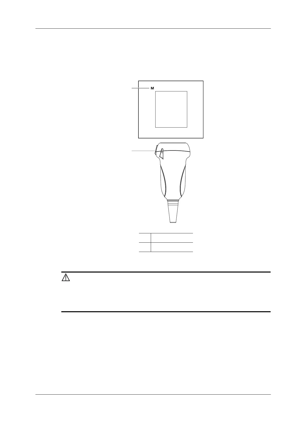
16.1.2 Orientation of the Ultrasound Image and the Probe
Head
The orientation of the ultrasound image and the probe are shown as below. The “M” side of the
ultrasound image on the monitor corresponds to the mark side of the probe. Check the orientation
before the examination (Using a linear probe as an example).
16.1.3 Procedures for Operating
Disinfect the probe and sterilize the needle-guided bracket before and after an
ultrasound-guided biopsy procedure is performed. Failure to do so may cause
the probe and the needle-guided bracket becomes a source of infection.
This section describes general procedures for operating the probe. The proper clinical technique to be
used for operating the probe should be selected on the basis of specialized training and clinical
experience.
1Orientation mark
2Mark
1
2
 Loading...