❯
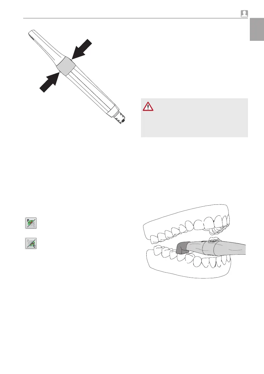
Press on one point of the capture ring.
The camera switches to "Freeze" mode. The
freeze frame will be displayed in the imaging pro-
gram / transmitted to the monitor.
❯
Edit the image using the imaging program and
save. (For further information, refer to the soft-
ware manual.)
❯
To return to "Live" mode, press on a point on
the capture ring again.
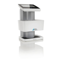
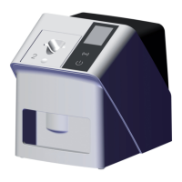
8.6 Record an image with the
Proof interchangeable head
When imaging with the Proof interchangeable
head, two views are possible in the imaging soft-
ware.
Prophylaxis view
This provides an informative overview of
the status of oral hygiene.
Caries view
It evaluates the fluorescence of the sub-
stances and provides a reliable diagno-
sis of carious lesions based on the
colours.
The following factors can affect the fluo-
rescence and hence the caries analysis:
–
Soiling and food remains
– Calculus, concrement
– Aid for staining plaque
– Prophylaxis/fluoride pastes
– Tooth/polishing pastes
Preparation
The teeth must be prepared differently depending
on the required analysis.
For prophylaxis view:
❯
Do not clean the teeth professionally.
For caries analysis:
❯
Carry out professional teeth cleaning.
❯
Remove polishing paste using the air/water
spray.
❯
Dry the teeth.
Record an image
CAUTION
The UV light of the camera can dazzle
❯
Do not peer into the light source.
❯
Do not use the camera directly on the
eye.
Requirements:
ü
Camera connected with the computer
ü
Imaging software started
ü
Camera in hygienic protective cover
ü
Spacer placed on
❯
Reduce the penetration of external light. Turn
off or dim sources of external light (e.g. operat-
ing lights).
❯
Place the camera with spacer onto the corre-
sponding tooth.
Usage
9000-618-176/30 1812V009 21
EN
 Loading...