8.7 Record an image with Proxi
interchangeable head
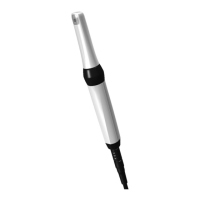
Positioning the unit correctly
The camera must be positioned correctly to
achieve a good picture quality.
❯
Position the camera in a line with the teeth.
❯
Place the spacer vertically on the tooth surface.
The spacer must come into contact with the
teeth.
❯
Ensure that the relevant approximal area is
located in the centre of the image section.
❯
If the structure underneath the enamel is not
visible, change the angle of the camera slightly.
Record an image
❯
Reduce the penetration of external light. Turn
off or dim sources of external light (e.g. operat-
ing lights).
❯
Dry the row of teeth with compressed air.
❯
Place the camera with spacer on the row of
teeth above the approximal area. The two
infrared LEDs illuminate the respective mesial
and distal enamel area of the two adjacent
teeth.
❯
Press on one point of the capture ring. The
camera switches to "Freeze" mode. The freeze
frame will be displayed in the imaging pro-
gram / transmitted to the monitor.
❯
Edit the image using the imaging program and
save. (For further information, refer to the soft-
ware manual.)
❯
To return to "Live" mode, press a point on the
capture ring again.
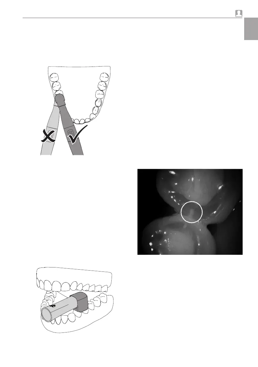
Analyse the image
The black and white image shows structures with
varying translucency as different levels of bright-
ness. The lower the translucency, the higher the
reflection of the infra-red light and the brighter the
structure. It is possible to make out to following
structures:
– Healthy enamel appears very dark, high
translucency
– Approximal caries appears bright, low translu-
cency
– Dentine appears bright, low translucency
– Several restorations appears bright, no translu-
cency
Fig. 10: Enamel lesions can be seen as wedge-
shaped structures within the dark translucent
tooth enamel. The lesions reach to the inner
half of the enamel.
The enamel appears brighter in patients with
highly opaque enamel. The caries diagnosis is
complicated here by the low difference in con-
trast.
Usage
9000-618-176/30 1812V009 23
EN
 Loading...