Chapter 2 Individual Operations
51
Chapter 2
Individual Operations
Light Reduction by Combined ND Filters of the Epi-fluorescence Attachment
Brightness ND4 ND8 ND16
1 - - -
1/4 ○ - -
1/8 - ○ -
1/16 - - ○
1/32 ○ ○ -
1/64 ○ - ○
1/128 - ○ ○
1/512 ○ ○ ○
(-: Removed from the optical path, ○: Placed into the optical path)
Improving the S/N ratio (shielding tube)
To improve the signal to noise ratio (SNR) during epi-fluorescence observations solely using epi-fluorescence
microscopy, we recommend removing the condenser and using the shielding tube provided with the epi-fluorescence
attachment.
In particular, when combining Ci-L and the epi-fluorescence attachment, excitation light from the Epi-fl attachment may
strike the white LED, making it illuminate and resulting in SNR deterioration. To avoid this situation, make use of the
shielding tube provided with the Epi-fl attachment or place a plate on the field lens.
■ ND on HG precentered fiber illuminator
You can also adjust the light intensity using the ND on the HG precentered fiber illuminator.
For detailed information, refer to the operating manual provided with the HG precentered fiber illuminator.
Locating a target on the specimen
The standard procedure for epi-fluorescence microscopy is to first locate the target under differential interference contrast
or phase contrast microscopy, and then switch to epi-fluorescence microscopy.
To locate the target under dia-illumination bright-field microscopy, you will need to note the following.
• Under dia-illumination bright-field microscopy, start with a 10x objective, and adequately stop down the condenser.
• Gradually increase the magnification. When the target becomes difficult to locate, switch to epi-fluorescence, and
use low excitation light.
• You can also use other techniques, such as using the edge of the cover glass to approximate the position of the
target.
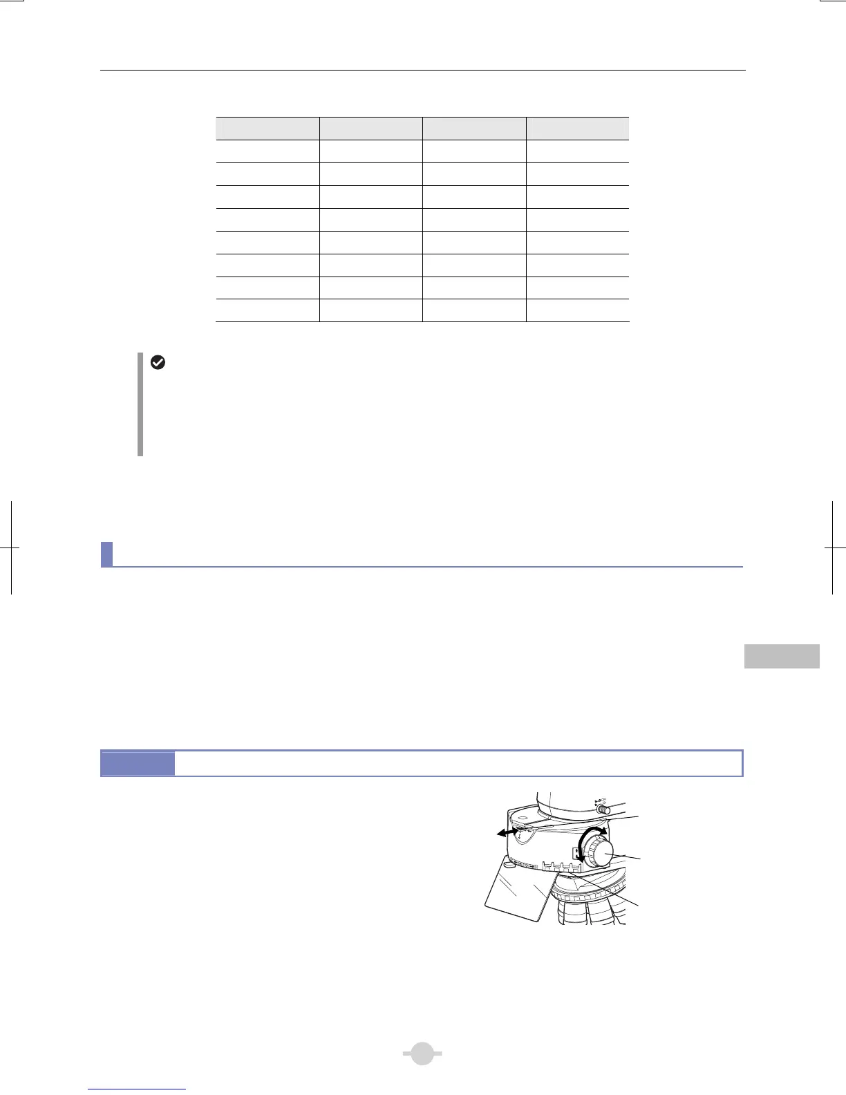
15.1
Switching Excitation Methods
Four filter cubes can be inserted into the epi-fluorescence
attachment. (→See Chapter 3, “6 Assembly for Epi-fluorescence
Microscopy”)
Move the desired cube into the optical path by turning the filter
cube switching knob on the right side of the attachment.
For bright-field observations, leave one cube position empty, and
move this empty position into the optical path.
Use the filter cube motion restricting lever located at the uppe
front section to limit the cube switching operation.
A1
-
2
-
3
-
4
B1
-
2 / 3
-
4
C
1
-
2
-
3
-
4
1
1
4
2
3
4
CUBE
Switching the filter cube
Filter cube
motion restricting
leve
Filter cube
switching knob
Filter cube
nameplate window
 Loading...