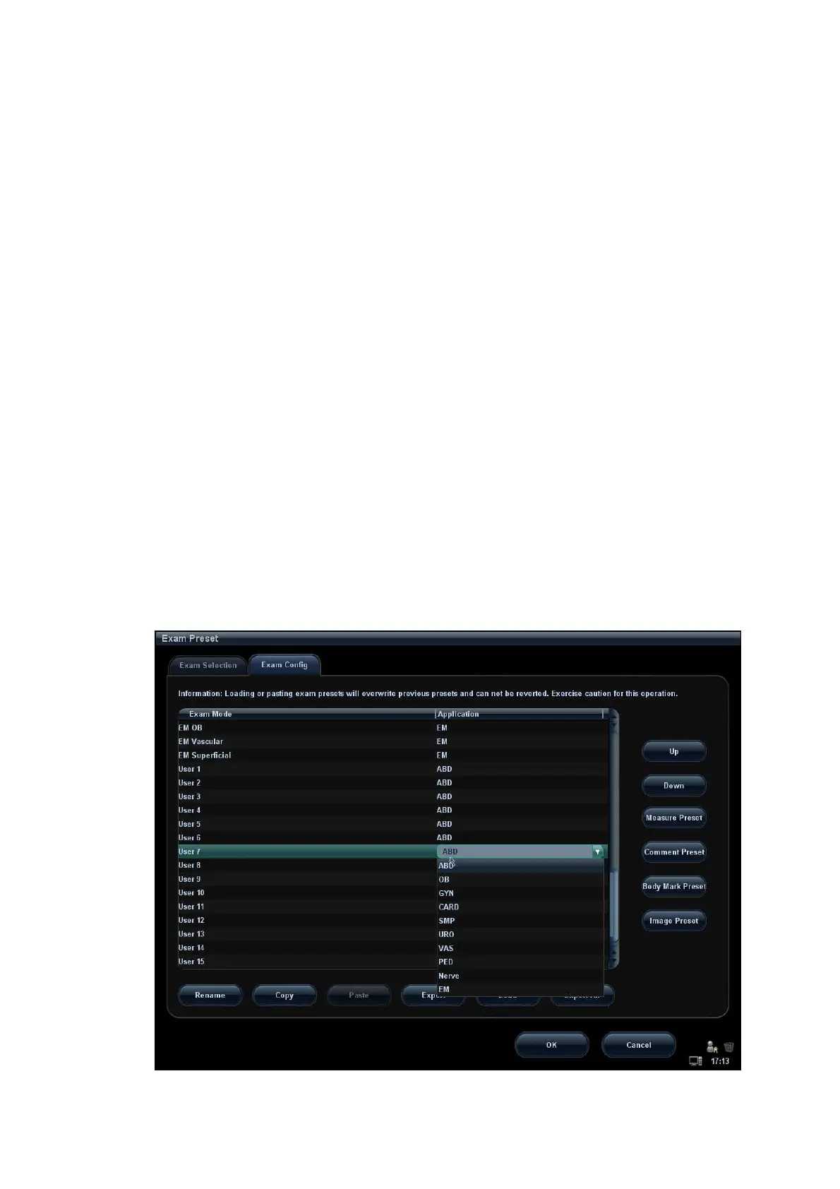
EasyManua.ls Logo

Mindray M6 User Manual

Mindray M6
357 pages
To Next Page IconTo Next Page
To Previous Page IconTo Previous Page
Page #300 background imageLoading...
Page #300 background image
14-12 Setup
After an exam mode is selected,
Click a selection in Application list, you can select a new application for the exam
mode in the popped up drop-down list.
Click [Up] and [Down] to adjust the sequence of the items in Selected Items.
Click [Rename] to change the name of the user-defined exam mode.[Rename]
Click [Copy] to copy the parameters of the selected exam mode. Select a second
exam mode and, click [Paste] to paste the copied parameters to the
secondly-selected exam mode.
Click [Export] to open the screen to store the parameters of the selected exam
modes.
Click [Load] to open the screen to load the parameters of the exam mode.
Click [Export All] to open the screen to store the parameters of all exam modes for the
current probe.
In addition, you can preset measurements, comments, body marks and image
parameters for the exam modes by clicking [Measure Preset], [Comment Preset],
[Body Mark Preset], [Image Preset] respectively.
Tips: loading or pasting exam setup data will overwrite previous presets and can't be
reverted. Exercise caution for this operation.
14.2.3 User-defined Exam Modes
To define exam modes for a probe:
1. Click to select a user-defined item in the [Exam Mode] list in Exam Config page, such
as [User-defined 1].
Click a selection in Application list, and select an application for the exam mode in the
popped up drop-down list (see the figure below).

Table of Contents

Questions and Answers:

Mindray M6 Specifications

General IconGeneral
TypePortable Ultrasound System
ApplicationsAbdominal, Cardiac, Vascular, Small Parts, Musculoskeletal
Operating SystemWindows-based
PortsEthernet, USB
Supported ProbesConvex, Linear, Phased Array
Imaging ModesB-mode, M-mode, Color Doppler, Power Doppler, Pulsed Wave Doppler, Continuous Wave Doppler

Summary

Safety Precautions

Safety Classification

Classifies equipment based on shock protection and environmental factors.

Meaning of Signal Words

Defines hazard indicators like DANGER, WARNING, CAUTION, NOTE, Tips.

Safety Precautions

Lists crucial safety guidelines for system operation and patient care.

Warning Labels

Explains system warning labels and their meanings for user safety.

System Overview

Intended Use

Defines the system's application scope and target users.

System Configuration

Details standard and optional system components available.

Control Panel

Provides a detailed description of all buttons and controls on the system panel.

System Preparation

Move/Posit the System

Instructions for safely positioning the ultrasound system unit.

Power ON /OFF

Describes the correct procedures for powering the system on and off.

Connecting /Disconnecting a Probe

Step-by-step guide for attaching and detaching ultrasound probes.

Exam Preparation

To Start an Exam

Outlines the general procedure for initiating a new patient examination.

Patient Information

Details how to enter and manage patient demographic data for exams.

Select an Exam Mode and Probe.

Guides the user through selecting appropriate exam modes and probes.

Pause Exam and End Exam

Explains how to pause and properly conclude an ongoing patient exam.

Image Optimization

Image Adjustment

Covers adjusting image parameters for optimal display quality.

B Mode Image Optimization

Specific settings for optimizing B-mode image quality.

3D/4D

Introduces advanced 3D and 4D imaging capabilities and usage.

Image Preset

Describes how to configure and save image parameters for different modes.

Display & Cine Review

Image Display

Covers basic image display features like splitting the screen.

Cine Review

Explains how to review recorded image loops and sequences.

ECG

ECG Operation Basic Procedures

Outlines the steps for connecting and operating the ECG module.

Measurement

Basic Operations

Covers fundamental procedures for performing measurements.

General Measurements

Lists various general measurement tools available for different imaging modes.

Comments and Body Marks

Comments (Annotations)

Details on adding text annotations to images.

Body Marks (Pictograms)

Instructions for adding anatomical pictograms to images.

Patient Data Management

Patient Information Management

Procedures for managing patient data.

Image File Management

Covers storing, reviewing, and analyzing image files.

Backing Up and Erasing Files through DVD Drive

Procedures for backing up system data to DVD.

Administration

Covers user access control and system settings.

DICOM

DICOM Preset

Configuration steps for DICOM connectivity.

DICOM Service

Overview of available DICOM services.

DICOM Storage

How to send images to a DICOM storage server.

DICOM Print

How to send images to a DICOM printer.

Probes and Biopsy

Probe

Information about supported ultrasound probes.

Probes Cleaning and Disinfection

Critical procedures for probe hygiene and maintenance.

Biopsy Guide

General safety and procedural information for biopsy.

Basic Procedures for Biopsy Guiding

Step-by-step guide for performing biopsy.

Verifying the Biopsy Guide Line

Essential step for accurate biopsy guidance.

Recording

DVR Recording

Procedure for recording examination videos.

DVR Video Replay

Procedure for replaying recorded videos.

Setup

System Preset

Configuring general system settings.

Key Config

Customizing control panel and footswitch functions.

Exam Preset

Setting up exam-specific parameters.

Batteries

Precautions

Safety guidelines for battery handling and usage.

Installing and Removing the Batteries

Procedures for battery installation and removal.

Acoustic Output

Concerns with Bioeffects

Information on potential bioeffects of ultrasound.

ALARA Principle (As Low As Reasonably Achievable)

Explains the principle of minimizing ultrasound exposure.

Acoustic Power Setting

How to adjust ultrasound output power.

Acoustic Power Control

Details on system controls affecting ultrasound output.

EMC Guidance and Manufacturer’s Declaration

TABLE 1 GUIDANCE AND MINDRAY DECLARATION—ELECTROMAGNETIC EMISSIONS

Information on electromagnetic emissions compliance.

System Maintenance

Daily Maintenance

Routine cleaning and checks for system upkeep.

Cleaning the System

Detailed instructions for cleaning system components.

Troubleshooting

Steps to resolve common system issues.

Appendix A Wireless LAN

To Use the W-LAN Function

Steps for configuring wireless network connectivity.

Appendix B To Use the Lock

To lock the machine

Procedure for securing the system with a lock.

Appendix C Barcode Reader

1-D Barcode Reader

Overview and setup of the 1D barcode reader.

2-D Barcode Reader (Take DS6707 as an example)

Overview and setup of the 2D barcode reader.

Setting in Ultrasound System

Integrating barcode reader with the ultrasound system.

Appendix D iScanHelper

Enter/ Exit

How to access and exit the iScanHelper feature.

Use iScanHelper for Reference

Using the feature for diagnostic reference.

Appendix E Electrical Safety Inspection

Power Cord Plug

Inspection criteria for the power plug and cord.

Protective Earth Resistance

Safety test for protective earth connection.

Earth Leakage Test

Electrical safety leakage test procedures.

Enclosure Leakage Test

Safety test for enclosure leakage.

Patient Leakage Current

Safety test for patient leakage current.

Mains on Applied Part Leakage

Safety test for applied part leakage.

Patient Auxiliary Current

Safety test for auxiliary current.

Related product manuals