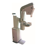
GE Healthcare Senographe DS Acquisition System
Revision 1 Operator Manual 5307907-3-S-1EN
Specifications
17-specs.fm Page no. 201 Chapter 17
2 Radiation and filter Information
2-1 Source to Image Distance (SID)
• Fixed SID: 660 mm
2-2 Radiation reference axis
Conforming to standard mammography practice, the radiation reference axis is directed at the chest wall
edge of the digital detector; radiation is shielded so that there is no radiation directed behind the chest
wall.
2-3 Leakage technique factors
With respect to radiation regulation, the tube housing and the collimator are in compliance with DHHS 21
CFR1020:
Leakage technique factors applicable: 49 kV at 2 mA.
2-4 Nominal X-ray tube voltages and currents
• Nominal X-ray tube voltage and the highest X-ray tube current available at that voltage:
49 kV; 61.2 mA
• Highest X-ray tube current and the highest X-ray tube voltage available at that current:
100 mA; 30 kV
• Corresponding combination of X-ray tube voltage and X-ray tube current which results in the highest
electrical output power (3 kW):
30 kV; 100 mA or 49 kV; 61.2 mA
• Nominal electric power given as the highest constant electric power (in kW) which the X-ray genera-
tor can deliver, for a loading time of 0.1 s at an X-ray tube voltage of 30 kV:
100 mA x 30 kV = 3 kW
2-5 Irradiation in AOP mode
• Nominal shortest irradiation time in AOP mode: 40 ms.
• Range of X-ray tube voltage during irradiation in AOP mode: 24 through 35 kVp.
• Range of X-ray tube current during irradiation in AOP mode: 30 through 100 mA.
2-6 Nominal focal spot size
• Large focal spot: 0.3 mm.
• Small focal spot: 0.1 mm.
FOR TRAINING PURPOSES ONLY!
NOTE: Once downloaded, this document is UNCONTROLLED, and therefore may not be the latest revision. Always confirm revision status against a validated source (ie CDL).
 Loading...