24
5
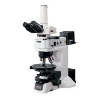
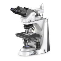
Sensitive Polarization Microscopy
Only when configured with the LV-UEPI2
6
x
6
STAG
E
JAPA
N
A
.
STOP
F
.
STOP
JAPAN
FL1
S
FL2
BF
DF
1
0
0
2
0
0
1
0
0
IN
O
U
T
L
V
-T
T
2
6
x
6
S
TA
G
E
J
A
P
A
N
A
.
STOP
F
.
STOP
JAP
AN
FL1
S
FL2
BF
DF
1
00 2
0
0
1
0
0
I
N
O
U
T
LV
-T
T
2
4
5
4
5
1
2
1
3
2
3
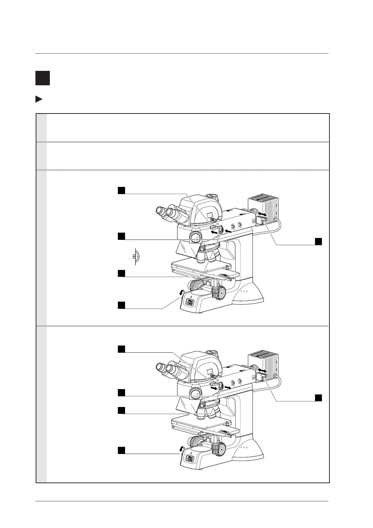
1. Mount a polarizer, lambda plate, and analyzer. (p.36 to 38)
2. Focus on the sample with bright-field microscopy. (p.17)
4. Return the microscope to bright-field microscopy.
Adjust the
brightness.
ND filter (p.26)
Adjust the
brightness.
ND filter (p.26)
Adjust the
brightness.
Brightness control
knob (p.26)
Adjust the
brightness.
Brightness control
knob (p.26)
3. Set the microscope for sensitivity polarization microscopy.
Push in.
Analyzer (p.38)
Pull out.
Lambda plate (p.37)
Pull out.
Analyzer (p.38)
Pull out.
Polarizer (p.36)
Push in and align
the marks.
Polarizer (p.36)
Crossed Nicols
position.
Information:
Turn the polarizer knob
to adjust the polarization
while observing the
image.
Push in.
Lambda plate (p.37)
 Loading...