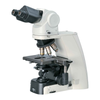
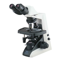
Chapter 1 Microscopy Procedures
34
Microscopy Procedures Epi-fluorescence Microscopy
Chap. 1-5
5.2
Epi-fluorescence Microscopy Procedure
WARNING
The light source used with the epi-fluorescence attachment (mercury lamp) requires special care during handling
because of its characteristics. Make sure you are familiar with and adhere to all warnings and cautions described at
the beginning of this instruction manual.
Locate the observation target on the specimen under bright-field microscopy, then proceed to epi-fluorescence
microscopy.
(See Chapter 2, “17 Tips for Epi-fluorescence Microscopy” for the tips to locate the observation target on the specimen.)
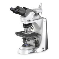
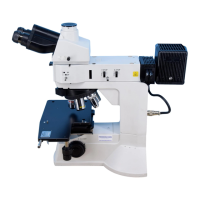
A CI-FL Epi-fluorescence attachment is used in the following procedure as an example. When using a D-FL
Epi-fluorescence attachment, refer to the D-FL Epi-fluorescence attachment Instructions.
1. Turn ON the dia-illumination LED.
2. Close the shutter.
3. Bring the filter cube into the optical
path.
4. Fully open the field diaphragm.
5. Turn on the mercury lamp.
6. Open the shutter.
7. Bring the desired objective into the
optical path.
8. Focus on the specimen.
9. Circumscribe the field diaphragm
to the field of view.
10. View the specimen.
11. Turn off the mercury lamp.
12. Turn off the power.
OFF 1 2
TOGGLE
PATTERN
MEMORY
CLAMP
TORQUE
0.1
0.3
0.4
0.5
0.2
0.9
0.8
INTENSILIGHT
C
-
HGFI
LAMP
ND
1
2
4
32
16
8
SHUTTER
POWER
RUN TIME hrs.
A1
-
2
-
3
-
4
B1
-
2 / 3
-
4
C1
-
2
-
3
-
4
1
1
4
2 3 4
CUBE
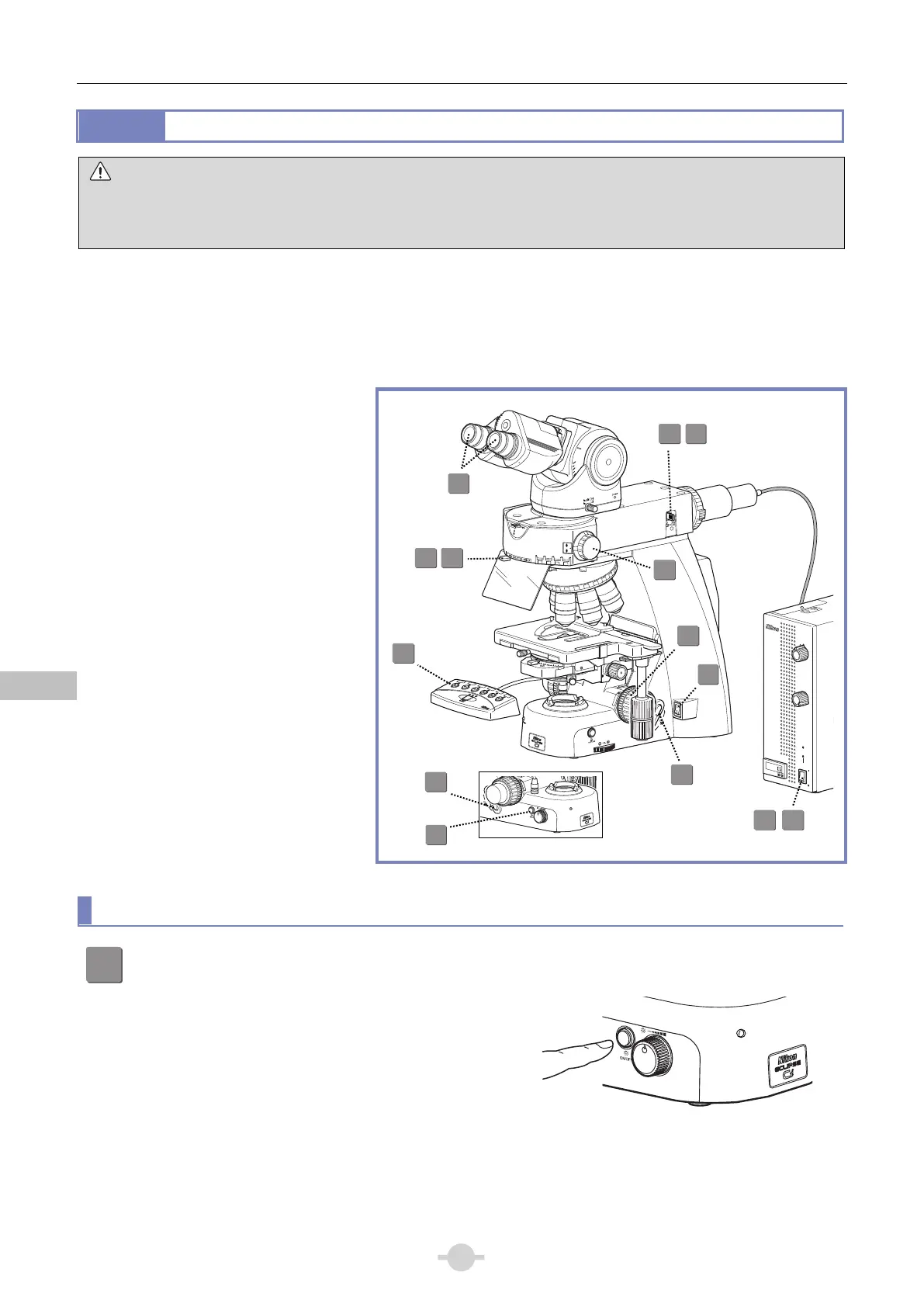
Microscopy operation (→See also: Chapter 2, “17 Tips for Epi-fluorescence Microscopy”)
1
Turn off the dia-illumination LED.
Press the dia-illumination LED ON/OFF switch to turn off
the LED.
P
O
WER
Dia-illumination LED switch OFF
8
10
3
2 6
1
7
5
11
9
4
TORQUE
C
P
O
WER
0
90
80
70
60
50
40
30
20
10
12
7
7
 Loading...