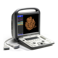
S6 Portable Digital Color Doppler Ultrasound System
Service Manual
through the computer system, the signals are sent to the graphics card and
then to the display (e.g. LCD monitor).
The computer system is the core control unit of S6. Following user in-
structions from the keyboard, it controls the operating modes and adjusts
the status of the system. The computer system is also responsible for the
display of diagnostic data, calendar, time and etc.
4.3 Principle of Probes
The transducer provides conversions between ultrasonic and electronic sig-
nals. At the start of a scanning process, the probe first converts the elec-
tronic excitation signals to ultrasonic vibrations. The ultrasonic vibrations
(or ultrasound) propagate into the body of the patient; and the echoes are
picked up by the probe. Electronic signals converted from these echoes are
sent back to the computer system or the DBF board for further processing.
The acousto-electric conversion efficiency and the ultrasound focusing abil-
ity are the key features that are vital to the system’s performance, such as
resolution, maximum scanning depth.
4.4 Operating Modes
This section gives brief explanations of the principles of the scanning modes
of the S6 system.
M Mode
Primarily applied in cardiology, M mode provides Time and Motion echo
information derived from a stationary ultrasound beam. It records moving
anatomical structures and produces subtle patterns of motion.
2D/B Mode
B mode image, also called 2D image, provides a cross sectional view of
tissues. The ultrasound image is derived from the tissue echoes that re-
ceived by the probe. Each echo’s intensity is mapped to a shade of gray;
and the location that the echo occurs is mapped to a unique point on the
screen. Except for linear array probes which produce rectangular images,
most probes produce fan shaped images.
Spectral Doppler(PW/CW)
The spectral Doppler mode detects the movements of red blood cells based
on the Doppler principle. The moving cells reflect the ultrasound sent by
the probe. The echoes are frequency shifted (phase shift are detected for
PW mode) as a result of Doppler Effect. The spectral distribution of the
echoes reveals details of the blood flow: red (blue) shift implies flow away
from (towards) the probe head. The pulsed wave mode (PW) is normally
applied at detecting low flow velocity, while the continuous wave mode
(CW) is normally for measuring high speed blood flow.
P/N: 4720-0034-01A
4-2
 Loading...