5-40 Image Optimization
a) Press <Set> to fix the rectangle's position, roll the trackball to change the size,
and press <Set> again to finish drawing the rectangle.
b) Move the cursor to the region you want to cut and press <Set> again to cut.
To undo the last cutting, Click [Undo] on the screen.
To undo all cuttings, Click [Undo All].
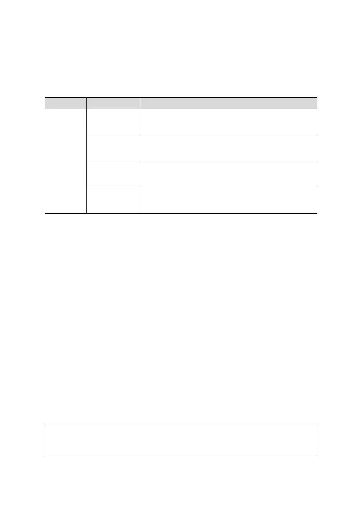
The image cutting parameters are described as follows:
Type Parameters Description
Edit Type
Inside Contour
Allows you to trace the portion of the image you want to cut.
Inside Contour removes all portions of the image that fall
inside your traced region.
Outside Contour
Allows you to trace the portion of the image you want to cut.
Outside Contour removes all portions of the image that fall
outside your traced region.
Inside Rect
Displays a box you can use to define the portion of the
image you want to cut. Inside Rect removes all portions of
the image that fall inside of the box.
Outside Rect
Displays a box you can use to define the portion of the
image you want to cut. Outside Rect removes all portions of
the image that fall outside of the box.
Section image (MPR) measurement.
2D related measurements can be performed on MPR. For details, see [Advanced Volume].
Measurement is not available in acquisition preparation status.
5.11.3.4 Image Saving and Reviewing in Static 3D
Image saving
In 3D viewing mode, press the single image Save key (Save Image to hard drive) to
save the current image to the patient information management system in the set
format and image size.
Save clip: in 3D viewing mode, press the user-defined Save key (Save Clip
(Retrospective) to hard drive) to save a CIN-format clip to the hard drive.
Image review
Open an image file to enter the image review mode. In this mode, you can perform the
same operations as in VR viewing mode.
5.11.4 Smart 3D
The operator manually moves the probe to change its position/angle when performing the
scan. After scanning, the system carries out image rendering automatically, then displays a
frame of the 3D image.
Smart 3D is an option. All probes for the Ultrasound System support the Smart 3D except for
D6-2EA.
5.11.4.1 Basic Procedures for Smart 3D Imaging
In Smart 3D image scanning, if the probe orientation mark is oriented to the
operator’s finger, perform the scan from right to left in linear scan, or rotate the
probe from left to right in rocked scanning. Otherwise, the VR direction will be
wrong.
To perform Smart 3D imaging:

 Loading...