S2/S2BW
Digital Color Doppler Ultrasound System
Chapter 9
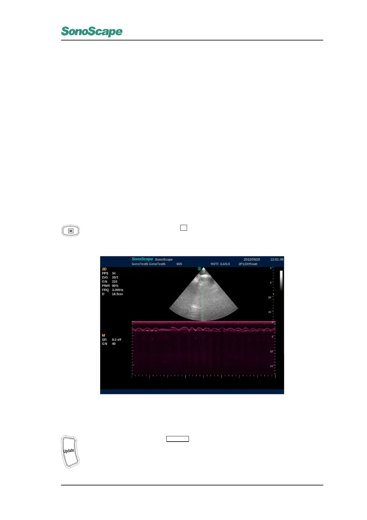
M Mode
M mode shows graph image about the tissue motion in a time sequence, the tissue motion information comes
from ultrasound beam echoes. M mode is mostly used in cardiology.
9.1 Starting M Mode
9.1.1 Pre-active Mode
When in B PW or CW mode, you press
M
, the image will divide into two parts, the upper one is the
B mode, the lower part of the screen is reserved for M mode graphs as the picture bellow shows. In
this picture the M mode is not really activiated, this is the “pre-active” mode:
Figure 9.1: M Mode Image
9.1.2 Active Mode
In the pre-active M mode, press
UPDATE
key to activate M mode. The M mode scanning graph
will show, now the upper part remains the B-mode image, and the lower part of the screen display
M mode spectrum.
P/N: 4710.00149A01
9-1
 Loading...