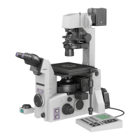
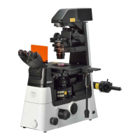
Chapter 2 Microscopy
25
2.5 Diascopic Dark-field (DF) Microscopy (Continued)
3
Adjust the condenser position to set up for DF
microscopy.
3-1 Stop down the dia field diaphragm.
[k] Field diaphragm dial (
☞
3.3.3)
▼
3-2 Raise the condenser until a dark shadow
appears in the field of view.
[o] Condenser focus knob (
☞
3.4.2)
▼
3-3 Move the black shadow to the center of the
field of view.
[t] Condenser centering screw (
☞
3.4.3)
▼
3-4 Adjust the height of the condenser to obtain
the highest contrast of the image.
[o] Condenser focus knob (
☞
3.4.2)
4
Perform DF microscopy.
4-1 Change the objective to the objective with a
specified magnification.
[b] Nosepiece (
☞
3.9.1)
▼
4-2 When using a liquid immersion type
objective, perform oil or water immersion.
[f] Objective (
☞
3.10.2, 3.10.3)
▼
4-3 When using an objective with a diaphragm,
align the diaphragm to an optimum position
below the minimum numerical aperture
of the condenser.
[f] Objective (
☞
3.10.4)
▼
4-4 When an objective with correction collar is
used, adjust the correction collar.
[f] Objective (
☞
3.10.1)
Adjust the correction collar of the objective according
to the thickness of the prepared specimen slide or the
cover glass of the culture vessel.
▼
4-5 Adjust the size of the dia field diaphragm
image to the field of view.
[k] Field diaphragm dial (
☞
3.3.3)
▼
4-6 Move the stage and turn the focus knobs to
focus on and observe the sample.
[m] Stage handle (
☞
3.8.3)
[q] Focus knob (
☞
3.11.1)
 Loading...