Eclipse Additional Information Page 122
5.11 Preparing for the oVEMP
5.11.1 Visual inspection of the ear canal
Check the external ear canal for wax with an otoscope and remove excessive wax to prevent the probe
opening from clogging which will inhibit testing.
Check if there might be middle ear issues, e.g. fluid or otosclerosis by performing tympanometry.
5.11.2 Preparation of the skin
Only apply the following procedure to patients for whom it is appropriate.
The electrode sites must be prepared and cleaned in order to obtain acceptably low skin impedance. A good
and thorough job of rubbing the skin with preparation gel may turn the skin a little red, but will ensure good
impedances. Some clinicians prefer to clean off the gel with an alcohol wipe. This will also ensure a very
clean area, well suited for the adhesive part of the electrode.
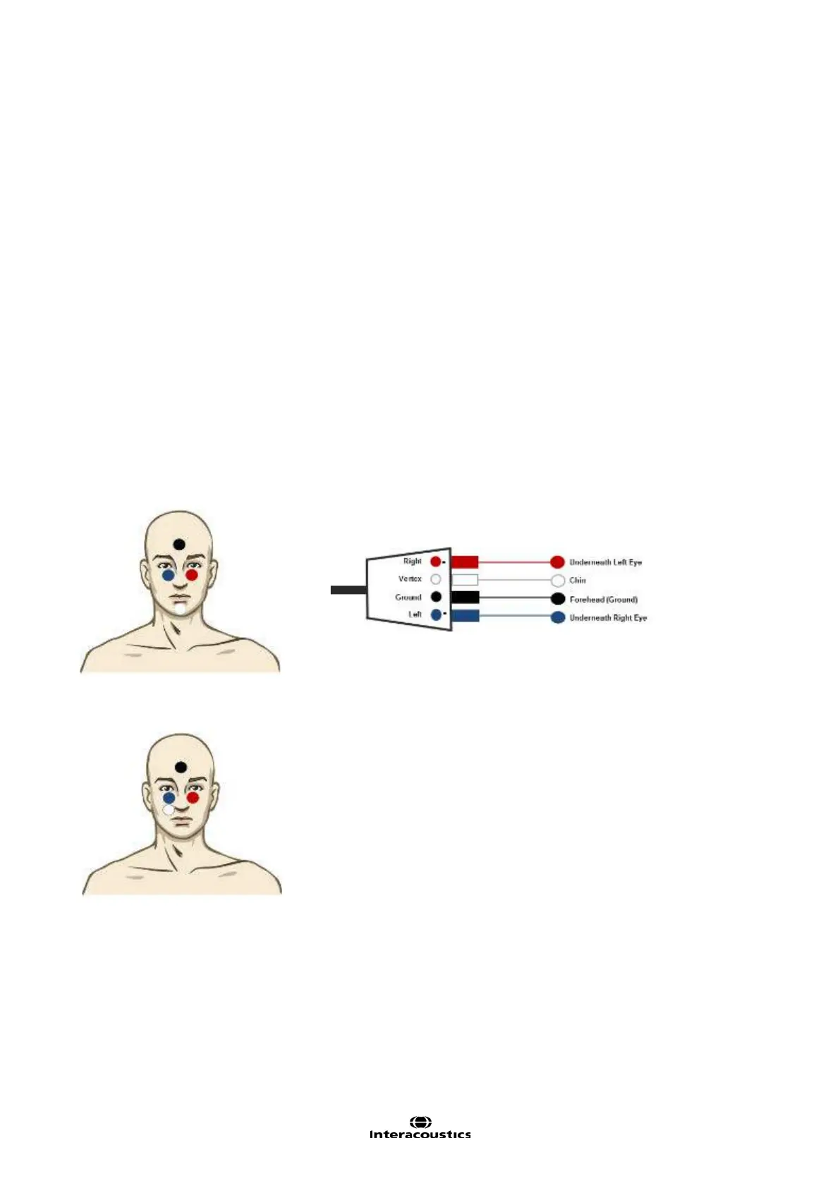
5.11.3 Electrode placement
The oVEMP response is recorded from the contralateral inferior oblique eye muscle. Therefore, the right
(red) electrode is placed on the inferior oblique muscle under the left eye while the right ear (utricle) is stimu-
lated. The reference electrodes should be placed as close as possible underneath the eye.
Alternative Electrode Placement
Electrode montage for testing left ear.
This electrode montage tends to create a greater oVEMP amplitude response. It is important to ensure that
there is a small distance between the red/blue electrode and the white electrode of approximately 1cm.
A disadvantage of this electrode placement is that the white electrode has to be moved to the opposite side
during testing. For the right ear testing, move the white electrode to other side (underneath the red elec-
trode).
Note The EPA3 cable collector does not allow VEMP testing, as the reduced VEMP gain cannot be enabled
on the preamplifier.
 Loading...