
Do you have a question about the Sonoscape E1 and is the answer not in the manual?
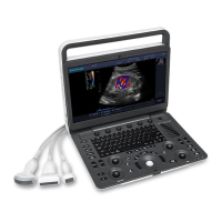
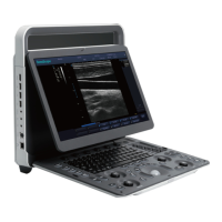
| Type | Portable Ultrasound System |
|---|---|
| Display | 15-inch LCD |
| Transducer Ports | 2 |
| Cine Loop | Yes |
| Imaging Modes | Color Doppler, Power Doppler |
| Applications | Obstetrics, Gynecology, Vascular, Musculoskeletal |
| Probe Connectors | 2 |
| Ports | VGA, USB, Ethernet |
| Supported Probes | Convex, Linear, Phased Array |
Outlines essential safety measures for system operation.
Details crucial safety guidelines related to electrical connections and usage.
Covers safety procedures for moving and handling the system physically.
Addresses safety protocols related to biohazards during system use.
Explains the principles governing ultrasound power output and safety.
Details the MI and TI indices for monitoring ultrasound safety.
Provides an overview of the various parts of the ultrasound system.
Explains the function and layout of the system's control panel buttons.
Details the functions of the keyboard and shortcut keys.
Describes the layout and elements of the main user interface screen.
Explains how to power the system using the adapter or battery.
Instructions for safely powering the system on, off, and into standby.
Procedures for properly connecting an ultrasound probe.
Configuration options for general system behavior and preferences.
Settings related to biopsy needle guides and guidelines.
Customizing and arranging exam presets for different probes and exams.
Adjusting parameters for various measurement functions.
Settings for DICOM connectivity and services like storage and printing.
Methods for entering and managing patient data for an examination.
Choosing the correct probe and application mode for imaging.
Capturing and optimizing standard B-mode ultrasound images.
Adjusting parameters like Gain, TGC, Focus, and Depth for optimal B-mode image quality.
Capturing and optimizing M-mode images for motion analysis.
Adjusting parameters like Gain, Chroma, and Gray Map for M-mode images.
Capturing and optimizing Doppler images for velocity analysis.
Steps to acquire Pulsed Wave Doppler images.
Adjusting parameters like Gain, Scale, Baseline, and Chroma for Doppler images.
Saving captured images and cine loops to the system.
Procedures for backing up system data to a USB drive or DICOM server.
Sending images and data to a DICOM storage server.
Configuring and sending the current image to DICOM storage.
Configuring and sending the current cine loop to DICOM storage.
Sending complete patient data records to the DICOM server.
Sending images for printing to a DICOM printer.
Guidelines for safely and correctly using ultrasound probes.
Procedures for cleaning ultrasound probes after use.
Methods for disinfecting and sterilizing probes according to required levels.
Specific methods for sterilizing probes used in critical applications.
Information and procedures related to using biopsy brackets and performing biopsies.
Step-by-step instructions for assembling biopsy brackets.
Detailed steps for performing a biopsy using the ultrasound system.
Scheduled checks to ensure system safety and functionality.
Guidance for resolving common system operational issues.