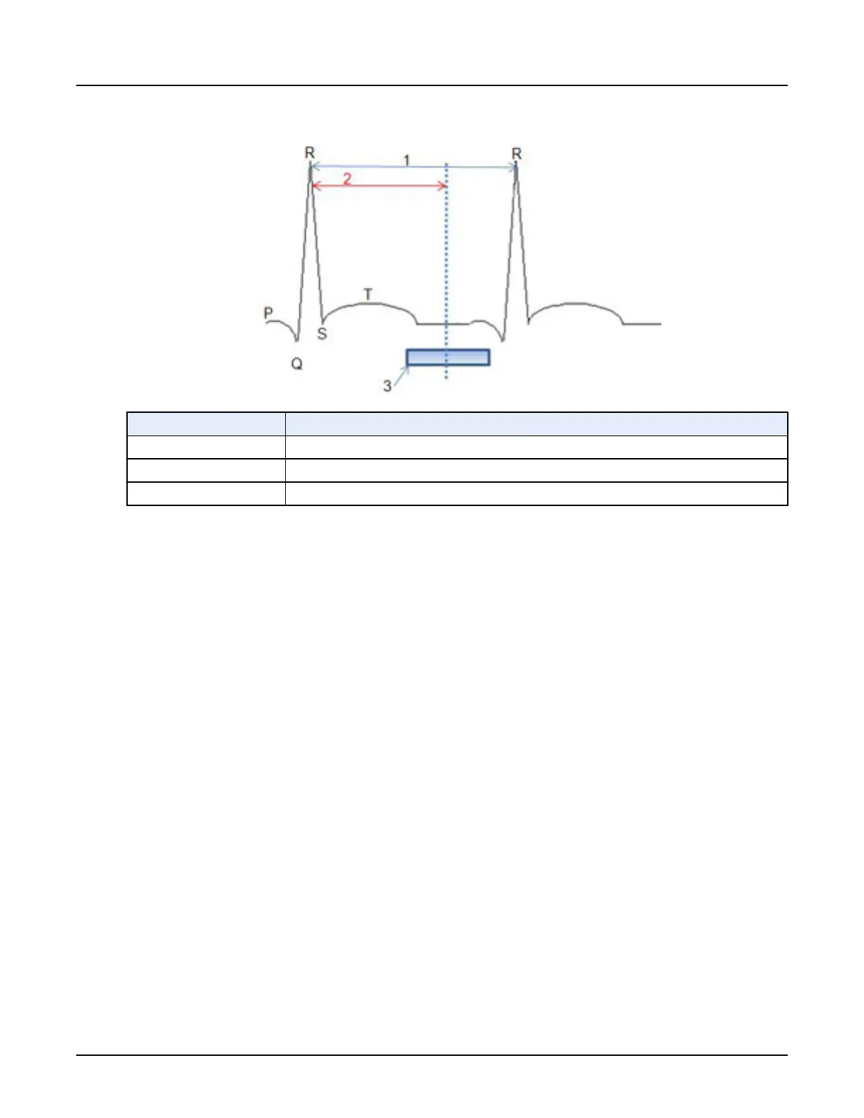
Illustration 11: ECG Waveform
Number Description
1 R-wave to R-wave interval
2 R-peak 75% default
3 Reconstruction window
•
Systole
, cardiac contraction, is from R-peak to T-wave.
•
Diastole
, cardiac relaxation, is from T-wave to R-peak.
R-wave to R-wave Interval
The R-wave to R-wave Interval is the time from the R-peak of one heart cycle to the R-peak of
the next heart cycle. The R-peak is used to predict where the targeted phase for reconstruction
occurs and then acquire/reconstruct images at that period of time during the heart cycle.
Phase Location (R-Peak Delay (%))
The cardiac phase location is defined as a period in time in the cardiac cycle. The Acquisition
Window Range setting (2) controls when the CT system will acquire data. Data is acquired so
that images can be reconstructed at all of the specified phases. (3) Refers to the center of the
reconstruction window in terms of a percentage distance between any two successive R-peaks
given from the ECG.
The cardiac phase location is defined as the time interval from the R-wave to the center of the
reconstruction window (3). It is usually desirable to reconstruct images at the phase that has the
least amount of cardiac motion. The phase location can be changed to identify where the least
amount of cardiac motion occurs. For lower heart rates, the most common location is in the
middle of diastole, typically around 75%, but as heart rate increases or varies this location may
change. It is not uncommon for higher heart rates to have better image quality at the end of
systolic portion, typically around 45%.
Revolution CT User Manual
Direction 5480385-1EN, Revision 1
352 3 Cardiac Scan Parameters
 Loading...