○
If you are acquiring a functional imaging exam for ejection fraction and wall motion,
prescribe phases from 5–95% in increments of 10% and consider reconstructing the
data with a 2.5 mm thickness.
12.
Click [Save] to close the Phase Selection dialog box.
NOTE:
A DICOM compliant ECG trace image is automatically saved after each primary and
secondary image reconstruction request. These traces are saved in a dedicated
Series labeled as
599
.
Use these images only as a record of the trace used to the generate images. The
images should not to be used for diagnostic purposes.
6.5 Interactive ECG Editor
Use these procedures to visually interact with cardiac reconstruction timing relative to the ECG
trace. This allows you to adjust gating information such as R-peak trigger time and
reconstruction timing relative to the ECG trace.
Considerations
If the accession number is not in the image header, it may cause the series to be listed as a
separate exam. If so, reconcile the exam on the PACS.
1.
Select a patient and exam for reconstruction and image processing; this will open the exam
on the image monitor.
2. Select the Series desired for reconstruction.
3. Make edits to R-peak triggers or reconstruction timing on the ECG trace.
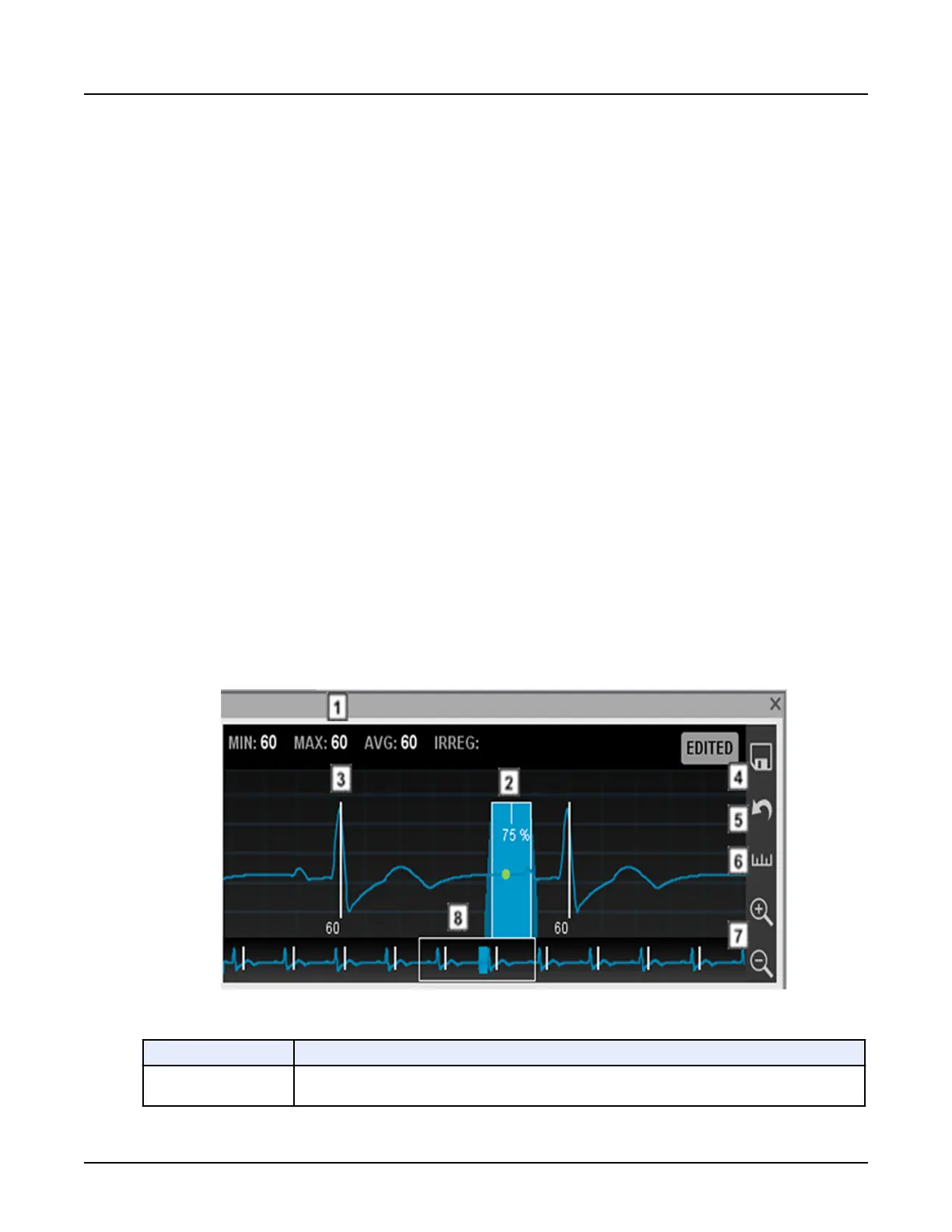
Illustration 15: ECG Editor window
Table 3: ECG Editor window
Number
Description
1
Heart rate statistics are displayed: minimum, maximum, average heart rates and number of irregular
beats.
Revolution CT User Manual
Direction 5480385-1EN, Revision 1
364 6 Cardiac Recon
 Loading...