Scanning Modes
Vivid S5/Vivid S6 User Manual 173
R2424458-100 Rev. 2
The Scale value also
affects the frame
rate. There is a trade
off between the
frame rate and
quantification
noise.
• To reduce quantification noise (variance), the Nyquist limit
should be as low as possible, without creating aliasing. To
reduce the Nyquist limit: Reduce the Scale value from the
assignables on the control panel.
PW will be opti-
mized for Tissue
Velocities when ac-
tivated from inside
TVI.
• TVI provides velocity information only in the beam direction.
The apical view typically provides the best window since the
beams are then approximately aligned to the longitudinal
direction of the myocardium (except near the apex). To
obtain radial or circumferential tissue velocities, a
parasternal view must be used. However, from this window
the beam cannot be aligned to the muscle for all the parts
of the ventricle.
Tissue Tracking
Tissue Tracking overview
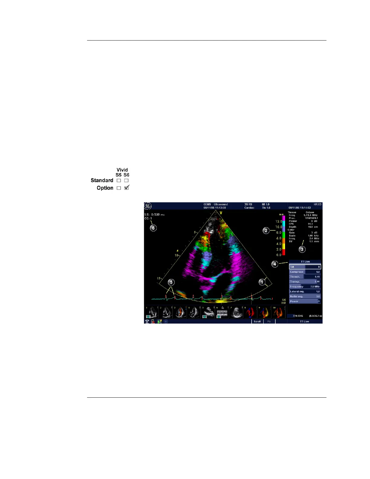
Figure 4-9: The Tissue Tracking Mode screen
1. Color sector marker
2. Tissue Tracking color bar
3. Status window
4. Soft menu
5. Track start and track end markers
6. Tracking start and end from R-peak

 Loading...