Preparation of slides Operation
27
4522 207 12671 * 2021-06-17
Pathology Scanner SG20 / SG60 / SG300
Preparation of slides
NOTICE
Prevent damage of the scanner
• Use only glass slides and cover slips supported by the scanner (see chapter “Glass slides,
cover slips and markers” on page 98).
• Make sure that the cover slip does not protrude over the edge of any portion of the glass
slide.
• Make sure that the label is positioned flat on the glass slide and does not stick out.
NOTICE
Improper tissue preparation can affect the image quality of the obtained image; e.g. tissue
folds can cause wrong focus and lead to image artifacts.
• Make sure that the tissue thickness is 3-5 µm.
• Make sure that the tissue is not folded.
• Make sure that the tissue is not over or under stained.
Follow the laboratory instructions to prepare the tissue, to mount it on the glass slide, to stain
the slide and to cover the slide with a cover slip. Follow the recommendations below for
optimal image quality and to minimize scan times.
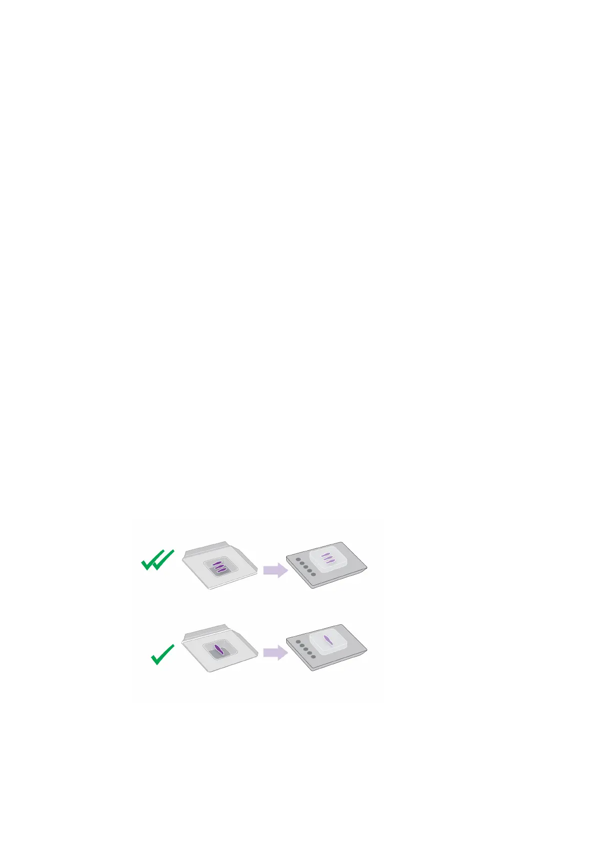
Preparing the block
► Keep in mind that the orientation of the tissue in the paraffin block has a small influence on
the scan time.
Fig. 10: Preparing the block
Cutting the tissue
► Use a microtome with a sharp blade to cut the tissue. Make sure that the tissue is
 Loading...