5-42 Image Optimization
Description of parameters:
Function: select the image acquisition method.
Selection: Rocked, Linear.
Linear mode: during the sweep, the probe must be kept parallel. The
scanning speed should be constant.
Rocked mode: in this mode, the probe must be moved to a position
where you can clearly see a middle cut of the object you want to scan
and render. Tilt the probe to about 30 degrees until the object you
want to scan disappears. Start the acquisition and tilt the probe over a
distance of around 60 degrees until the object disappears again.
During the sweep, the probe may not be moved parallel, just tilted.
Tip: the speed is related to scanning distance or angle.
Function: to set the distance the probe covered from one end to the
other end during a linear sweep.
Range: 10-200 mm, in increments of 10 mm.
Function: to set the angle the probe covered during a fan sweep.
Range: 10-80°, in increments of 2°.
Function: set Surface as the 3D image rendering mode.
This is useful for surface imaging, such as fetus face, hand or foot.
Tip: you may have to adjust the threshold to obtain a clear body
boundary.
Function: set Max. as the 3D image rendering mode. Displays the
maximum echo intensity in the observation direction.
This is useful for viewing bony structures.
Function: set Min. as the 3D image rendering mode. Displays the
minimum echo intensity in the observation direction.
This is useful for viewing vessels and hollow structures.
Function: set X-ray as the 3D image rendering mode. Displays the
average value of all gray values in the ROI.
X Ray: used for imaging tissues with different internal structures or
tissues with tumors.
5.11.2.3 Smart 3D Image Viewing
Enter/Exit Image Viewing
To enter image viewing:
The system enters image viewing when image acquisition is complete.
Exit
To return to Smart 3D image acquisition preparation status, tap [Update] or [Freeze].
Activate MPR
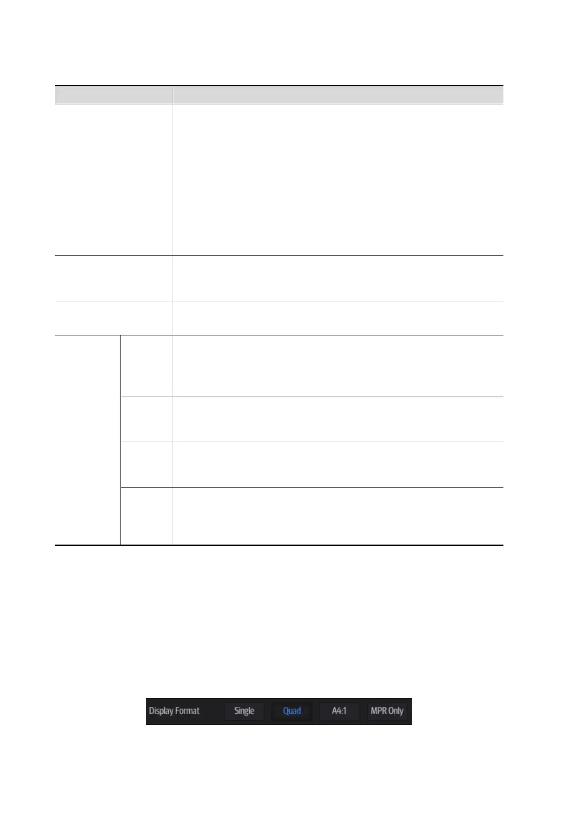
Tap [A], [B], [C] or [VR] to activate MPR or 3D image (VR).
 Loading...