Using the M4 TORNADO Software
134
User Manual
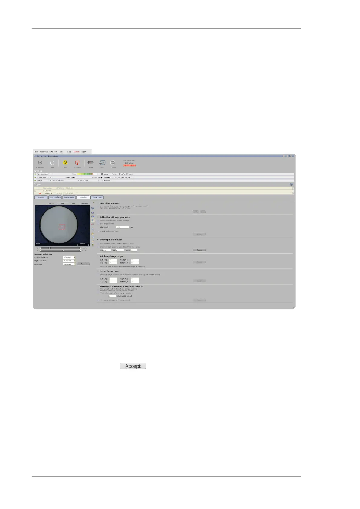
6.12.6 Settings for the X-ray spot calibration
A fine adjustment of the X-ray spot position to the sample image is possible by digital tuning of the
cross hair position in the working space System>>Imaging. The X-ray foil on the blue reference
sample should be positioned in the chamber and focused to the correct height. By activating the
menu point X-ray spot calibration the shutter opens at the current position of the X-ray spot
what can be seen in the video image. The fluorescence is produced from the Gadolinium foil. In
case the spot is not easy to recognize the illumination has to be reduced (mainly for collimators).
Tune the X and Y values to match the cross hair and the X-ray spot position for the 10x
magnification and repeat this for the 100x magnification. Note that the collimators are usually
> 1 mm and cover the complete 100x magnification image check location in the 10x magnification
image. In case of double excitation only the lens can be adjusted and the collimator position is set
at the factory.
Fig. 65 Adjustment of the X-ray spot position
The display of the spot size (red circle) can be adjusted to the real spot size of the optic with the
value in d/µm. Note that this value is defined by the manufacturer and does not change.
Settings must be confirmed with .
6.12.7 Settings in the area for autofocus calculation
The autofocus function will be done by the calculation of the contrast value of the selected image
region. The principle is based in the assumption that the focal plane has the highest contrast
value. In case of non-regular shaped samples it can happen that wrong parts of the sample will be
used for the autofocus. Therefore, the area used to perform the contrast calculation for the
autofocus can be set in System >> Imaging. The selected area will be displayed. It should be
only positioned around the spot position so that for non-regular shaped samples the autofocus
function works more reliable.
 Loading...