The same applies for the Threshold Volume result window see
(chapter
'Threshold Volume'
on
page 9-130
)
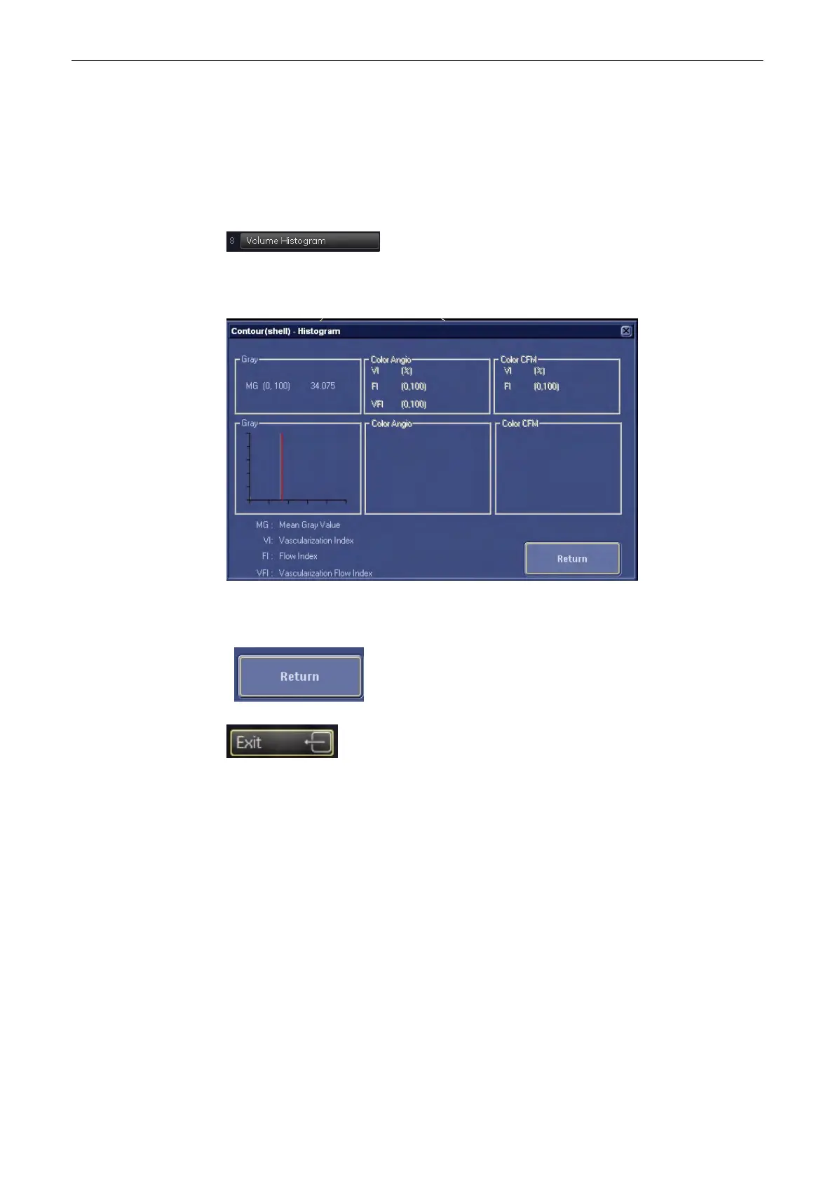
9.12.5.8 Volume Histogram
After volume calculation via the VOCAL program, it is possible to display an automatically
calculated (Color Angio) Histogram of the volume.
Select the [Volume Histogram] key in the Vocal menu.
Following window with the calculated histogram appears on the screen.
If a
shell
is defined, the histogram is calculated from the content of the shell. If a
contour
without a shell is defined, the histogram is calculated from the content of the contour.
Select the [Return] key or the [Exit] key on the menu area to exit the Volume Histogram
function.
Note
The Volume Histogram is not possible after 3D+CFM acquisition.
9.13 SonoAVC Follicle
General: SonoAVC Follicle is an option. If this option is not installed, the [SonoAVC Follicle]
key is hidden.
9.13.1 General Information
This feature automatically detects low echogenic objects (eg. follicles) in an organ (eg. ovary)
and analyzes their shape and volume. From the calculated volume of the object an average
diameter will be calculated. All objects detected that way will be listed according to size.
Volume Mode
Voluson® S6/S8 Basic User Manual
5433669-100 Revision 4 9-131
 Loading...