Home
Ziehm Imaging, Inc.
Science Education products
QUANTUM
Ziehm Imaging, Inc. QUANTUM User Manual
5
of 1
of 1 rating
220 pages
Give review
Manual
Specs
To Next Page
To Next Page
To Previous Page
To Previous Page
Loading...
Section 11.0
MAN 06–0017H
ZIEHM QUANTUM
Us
er’s Manual
Page 174 of 220
11.2
SHIPPING LOCKS INSTALLATION
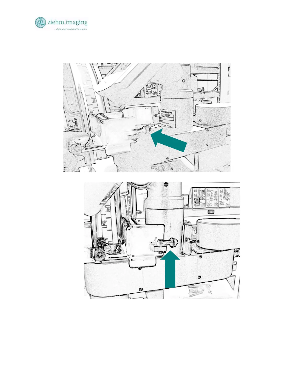
•
Push the shipping lock forward and while
simultaneously pulling the curved portio
n
of the bracket around the
DeskView support shaft. Example Fig, 11.10.
•
Tighten the locking bolt to secure
the shipping lock. Example Fig 11.11
173
175
Table of Contents
Default Chapter
3
Table of Contents
3
About this Manual
11
1 Safety & Responsibilities
13
Intended Use
13
Operation (U.s
13
Authorized Personnel
13
Equipment Owner Responsibilities
14
Hospital Administration
14
Manufacturer
14
Exclusion of Liability
14
Usa Regulatory Responsibilities
15
Service Engineers/Technician
15
System Users
15
Cdrh Report
15
Problems
15
Regulatory /Notified Body
15
Record Keeping Usa
15
2 System Overview Section
17
Fields of Application
17
Features
17
Mobility
17
Organ Programs
17
Radiation Reduction
17
Image Quality
17
Image Adjustment
17
Image Management
18
Documentation and Output
18
Networkability
18
Options
18
Optional Accessories
19
Parts of the System
20
C-Arm Stand
20
Twin Flat Panel Monitors
20
Monitor Position
21
Dual Flat Panel Lcd Monitors
23
Changing the Monitor Settings
24
Ziehm Quantum Fp Monitors Adjustments
24
Monitor Settings
24
Integrated Button Panel
24
Setting the Brightness, Contrast and Backlight Brightness
24
Setting the Menu Language
26
Restoring the Factory Settings
27
Bnc Socket Video Output
28
3 Safety Instructions
29
General Safety Instructions
29
Operation
29
Operation (U.s.a.) Assembly and Service
29
Warranty Voided
29
X-Ray and Electrical Safety
29
General Radiation Safety
29
Hazards
30
X-Ray Tube Housing
30
X-Ray Tube
30
Radiation Warning
30
Protection-Of Staff
30
Flammable Gas
31
Mechanical and Electrical Warnings
31
Electrical Safety
31
Grounding
31
High Voltage Components
31
Performing Internal Service
32
General Safety
32
Gettering
33
Bonding
33
Protection of the Patient
33
Electromagnetic Compatibility
34
Protective Earthing
34
Equipotential Earthing Heart and Brain Examinations
34
Laser Radiation
34
Laser Targeting Device
34
Maintenance
35
Laser Radiation
35
Environmental Compatibility
36
4 Unpacking Installation
37
Installation Procedure
37
Audience
37
Detailed Information
37
Room Temperature
37
Shipping Container Unpacking
37
Unpacking
37
Mounting Monitors
39
Mounting Standard Monitors
40
Mounting Quick Release Monitors
40
Power Cable Connections
42
Preparing the System
42
To Prepare the Unit, Do the Following
42
Switching on the System
43
Customer-Specific Initial Settings
43
Configuration
44
Entering the Hospital Data
44
Test Operation
44
Check Leakage Current
44
C-Arm Operation
44
Fluoroscopy Is Selected
44
Perform the Following Operational Tests & Verify that the Following Results Occur
45
Accessories
46
5 Mechanical Handling
47
Transport Position
47
Transport Position of the C-Arm Mobile Stand
47
Transport Position for Ziehm Quantum
51
Braking and Steering the C-Arm Stand
52
Steering & Braking
52
Parking Brake
52
Steering
52
Movement of the C-Arm, Mechanical Brakes
53
Orbital Rotation
54
Angulation
55
Swiveling (Panning)
55
Horizontal Movement
56
Vertical Movement
57
Vertical Override
57
6 User Touch and Console Keyboards
59
Deskview Tm Control Panel
59
Deskview Control Panel
59
Elements of the Control Panel
59
Deskview Main Screen Buttons
60
Main Screen Button Identification Table
61
Controls in Fluoroscopic Operation Main Screen
72
Touch Control Keyboard
72
Alphanumeric Keypad
73
Uppercase Letters and Special Characters
73
Mobile Stand Control Keyboard
74
Main Keyboard Element Groups
74
Mobile Stand Control Keyboard Table
75
Switching on the Ziehm Quantum
81
Switching off the Ziehm Quantum
82
Vertical Lift
83
Vertical Override
83
Hand Switch and Foot Switch
83
7 Standard Fluoroscopic Operations
85
General Description
85
Special Features
85
Image System Design
85
Imaging Processing
85
Thumbnail Image Display
86
Live Images
86
Fluoroscopy
86
Basic Operation
87
Steps and Settings
87
Adjustments that Can be Made
87
Fluoroscopy Modes
88
Continuous and Pulse Fluoroscopy
88
Continuous Fluoroscopy
89
Pulse Fluoroscopy
89
Snapshot
90
Fluoroscopy Anatomical Programs
90
Activating Anatomical/Organ Program
90
Bone/Extremity
91
Pelvis/Abdomen
91
Heart/Thorax
91
Lpd Large Patient Adipose Patient Function
91
Digital Noise Filters
92
Noise Filter Factors
92
Last Image Hold Noise Filter
92
Metal Artifact Correction
92
Manual Exposure Rate Setting
92
Screen Display During Radiation
94
Fluoroscopic
94
Fluoroscopy Factors
94
8 User Functionality
97
Special Functionality
97
Metal
97
1/2 Dose
97
Magnification Electro-Optical Image Intensifier
97
Mobile Stand Control Panel
97
Deskview Touch Panel
97
Image Rotation
98
Image Reversal / Image Mirror
98
Iris Collimator
98
Slot Collimator
98
Mark Image
99
Live Swap
99
Monitor L to
100
Monitor R to
100
Inv Video
100
Storing/Saving
101
Store Usb Image
102
Mobile Stand and Touch Information Displays
103
Kv Display
104
Ma Display
104
Exposure Time
104
Cgycm2 / Error
104
Min Fluoroscopy Time Display
104
Audible Alarm
104
Generator Overheating
105
Radiation Exposure Indicator
105
Deskview Displays for Kv/Ma, Time and Dose
106
Sor Seconds
106
Min
106
Mgy / Mgy/Min: (Active Only on System Manufactured after June 10, 2006)
107
Laser Aiming Device
107
Range of Applications
107
Positioning Aid
108
Dose Display for Akr and Accumulated Air Kerma
108
AKR /Cumulative Air Kerma Dose Display (Active Only on System Manufactured after June 10, 2006)
108
Display Functions
108
Dose Display User/Operator
108
Reset Dose Display
109
Image Filters
110
Rte (Real-Time Edge Enhancement)
110
Noise Reduction
110
Lih Noise (Last Image Hold Noise)
111
Auto Functions
111
Setting Auto Store
111
Auto off
112
Auto Store
112
Auto Transfer
112
Exit Auto
112
Post Processing Functions
112
W/L (Window Level)
113
Adjusting Window
113
Adjusting Level
113
Using the Window/Level Function
113
Invert
114
Image Crop
115
Zoom (Digital Magnification)
116
Selecting Thumbnail Images
118
Thumbnails
118
To View or Select a Specific Images
118
Store to or Make a Copy of an Image
119
Monitor Image Displays
121
Direct Radiography Option
122
Overview
122
Fitting the Film Cassette Holder
122
Manual Settings
122
Making a Direct Radiographic Exposure (Optional)
122
Set Technique Factors
124
9 Cine & Digital Subtraction Mode
125
Dsa/Cine Functions
125
Cine and Dsa Navigation Buttons
127
Performing Dsa
127
Storing of Dsa/Cine Images
128
Storing Individual Images to the Patient File Directory
129
Cine Functions
129
Cine Messages
129
Image Stop/Play and Step
130
Performing Cine Acquisition
130
Acquiring a New Cine Run
131
Image Step
131
10 Patient File Operations
133
Patient File Overview
133
New Pat (New Patient)
135
New Patient Entry
135
Patient Name
135
Facility Name
136
Escape (Esc)
136
Name Folder
136
Function
136
Storage Capacity
136
Edit Patient Records
137
Deleting Patient Records
139
Deleting Patients to Free up Space
140
Protecting a Patient File
141
Log in Security Permission
142
Image Export
143
Overview of the Usb and DVD Export Functions
144
Enter Archive
146
Image Archive Screen Button Description
147
Selecting Image Format
151
Select Patient Images for Export
152
Usb Memory Device Problems (Thumb Drive)
153
Optional: Send Dicom Images to the Pac's Server
154
Patient Folders
155
Configuring System: (Config)
156
Erasing Disk and Activating Database after Erase Disk Operation
156
Setting User/Administrative Log in Permissions
161
Restrictions
161
Setting User Permissions
163
Delete Single Log in Permission
163
Delete All Permissions
164
Save and Clear Export Log Files
164
Setting Time and Date
166
Setup Service Key and Service Log
167
11 Ziehm Quantum Optional Transport Brackets
169
Floor Mounted Installation
169
Shipping Locks Installation
174
Shipping Locks Removal
178
Floor Mounted Removal
178
Monitor Transport Container
179
12 Technical Data
181
Safety Classification
181
Statement of Compliance Iec Standards
181
Fda Product Classification Name / Code
181
Technical Specification
181
X-Ray Generator X-Ray Generator, Housing and Tube with Beam- Limiting Device
183
Mechanics
184
C-Arm Overall Dimension
186
Conversion Table MM to Inches
186
Scattered Radiation in the Significant Zone of Occupancy
187
Laser Alignment
188
Dose Akr and Cumulative Air Kerma Measurement
188
Calibration of Calculated Dose Measurement 21Cfr 1020.33
188
Akr / Cumulative Dose Display Calculation
188
Test Interval
189
Operator Monthly Test Procedure
189
Test Results
190
Dose Calibration Point
190
Heat/Cooling Capacity
192
Anatomical Program Kv/Ma Dose Curves
192
13 Inspection & Maintenance
197
Routine Checks to be Performed by the User
197
System Inspection
197
Table of Regular Checks
197
Test Recording
197
Verify Automatic Dose Rate Control
198
Checking the Useful Beam
198
Inspection Interval
198
Centering
198
Collimator Diameter Accuracy
198
Gettering the Image Intensifier Tube
199
To Getter the Image Intensifier Tube, Do the Following
199
Preventative Maintenance
199
Cdrh Maintenance
200
14 Cleaning, Disinfection, Sterilization
201
Preparation
201
Cleaning
201
Recommended Detergents
201
Cleaning the Monitor Screens
201
Disinfection Sterilization
201
Recommended Disinfectants
201
Alcohol or Sagrotan
201
Disinfecting Sprays
201
15 Appendix Labels
203
16 Appendix Error Codes
215
Error and Alert Messages
215
Errors During Normal Use
215
17 Glossary
217
5
Based on 1 rating
Ask a question
Give review
Questions and Answers:
Need help?
Do you have a question about the Ziehm Imaging, Inc. QUANTUM and is the answer not in the manual?
Ask a question
Ziehm Imaging, Inc. QUANTUM Specifications
General
Brand
Ziehm Imaging, Inc.
Model
QUANTUM
Category
Science Education products
Language
English
 Loading...