6 - 22 Operator’s Manual
6 Image Acquisition
• It supports single-frame and multi-frame image file detection in B mode.
6.10.1 Basic Procedures for Smart B-line
Perform the following procedure:
1. Select an appropriate probe and exam mode. The system enters the B mode by default.
2. Adjust the image parameters to obtain optimized images.
3. Select [Smart] > [Smart B-line] to enter Smart B-line mode.
Select the different zone combinations for examination.
4. Select a desired zone, and select the [Auto Calc] button.
The system automatically starts tracing the B line sampling area, and automatically recognizes
and traces the B line in frame.
If necessary, you can adjust the B line sampling area: Tap the dotted circle and drag the
sampling line to change the position.
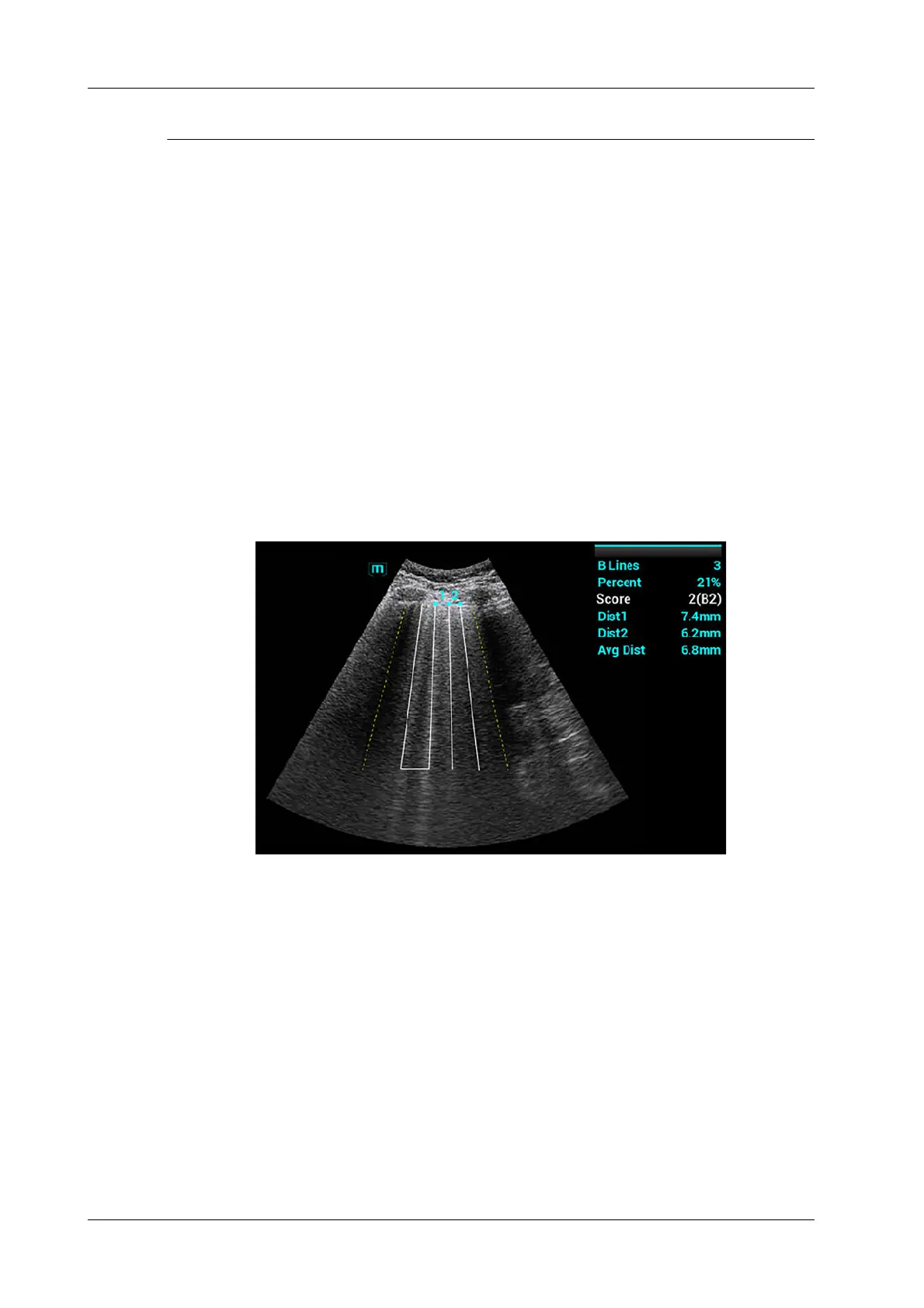
5. Tap [Freeze] button to freeze the image.
The system automatically calculates the quantitative index, and the calculation results are
displayed on the screen.
– B Lines: indicates the number of B lines of the current frame. The number can be 1, 2, 3,
4, or ≥5. When the number is equal to or greater than 5, the system does not display a
specific number.
– Percent: indicates the percent of the B lines area against the total sampling area.
– Score: the score is among 0 to 3.
Normal: when there are a lung sliding sign and A line, or isolated B lines (<3), it is
marked as N in the brackets and the score is 0.
Moderate: when there are multiple clearly-distributed B lines, it is marked as B1 in the
brackets and the score is 1.
Severe: when there are intensively fused B lines, it is marked as B2 in the brackets and the
score is 2.
Lung consolidation: when the lung has a symptom that is similar to the liver lesion
structure and air bronchogram, it is marked as C in the brackets and the score is 3. When
 Loading...


