8 - 4 Operator’s Manual
8 Smart 3D
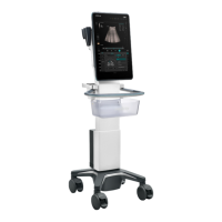
• Section A: corresponds to the 2D image in B mode.Section A is the sagittal section in fetal face
up posture, as shown in the figure A above.
• Section B: it is the horizontal section in fetal face up posture, as shown in the figure B above.
• Section C: it is the coronal section in fetal face up posture, as shown in the figure C above.
The ultrasound images are provided for reference only, not for confirming a
diagnosis. Please use caution to avoid misdiagnosis.
8.2 Note before Use
The quality of images reconstructed in the freehand 3D mode is closely related to the fetal
condition, angle of a B tangent plane and scanning technique. The following Smart 3D description
uses the fetal face imaging as an example, the other parts imaging are same as 3D imaging.
• In accordance with the ALARA (As Low As Reasonably Achievable) principle, please try to
short the sweeping time after a good 3D imaging is obtained.
• A region with qualified image in B mode may not be optimal for 3D imaging. E.g. adequate
AF isolation for one section plane of 2D image doesn’t mean the whole desired region is
isolated by AF for 3D imaging.
• More practices are needed for a high success of qualified 3D imaging.
• Even with good imaging condition, to acquire an approving 3D image may need more than one
scanning.
Fetal Condition
• Gestational age
Fetuses of 24~30 weeks old are the most appropriate to 3D imaging.
• Fetal body posture
Recommended: Cephalic face up (figure a) or face aside (figure b); NOT recommended:
Cephalic face down (figure c).
 Loading...


