13 Probes and Biopsy
Operator’s Manual 13 - 21
The biopsy needle may not have actually entered the target object even
though it appears to have done so in the image. To avoid this, note the
points below:
– Do not rely only on the needle tip in the image. Pay careful attention to
the fact that when the biopsy needle enters the target object or comes
into contact with it, the object should shift slightly.
– Before performing the biopsy, evaluate the size of the object and
confirm whether the biopsy can be carried out.
13.2.1 Needle-guided Brackets Available
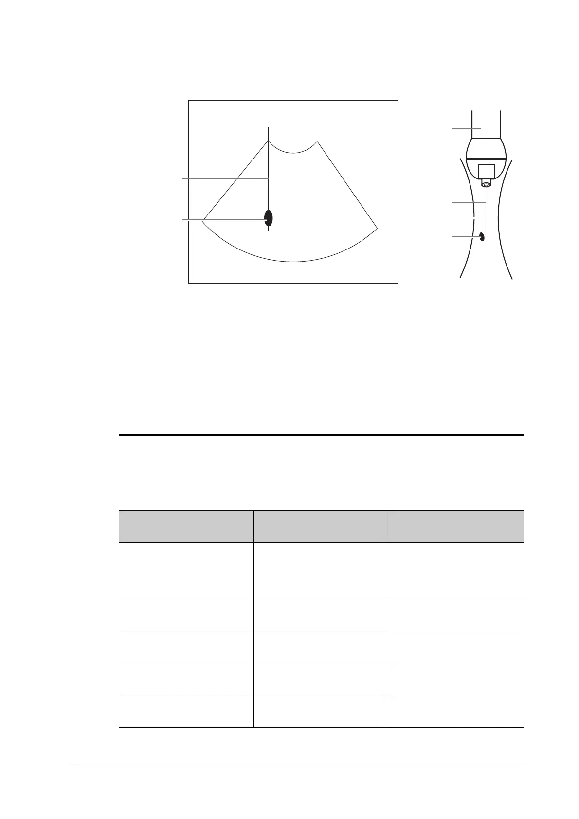
Probe
Needle
Ultrasound
beam
Targe t
Biopsy
Targe t
The biopsy needle appears to reach the target object
in the image
Dispersion of the
ultrasound beam
Table 13-4 Needle-guided Brackets Available
Needle-guided Bracket
Model
Biopsy angle/depth
(±1°)
Applicable Biopsy Needle
NGB-007
plastic/needle detachable
metal/needle detachable
40°, 50°, 60° Metal: 14G, 16G, 18G, 20G,
22G
Plastic: 13G, 15G, 16G, 18G,
20G
NGB-010
metal-needle detachable
30°, 40°, 50° 13G, 15G, 16G, 18G, 20G
NGB-011
metal/needle undetachable
11°, 23° 13G, 15G, 16G, 18G, 20G
NGB-016
metal/needle detachable
30°, 40°, 50° 14G, 16G, 18G, 20G, 22G
NGB-018
metal/needle detachable
15°, 25°, 35° 14G, 16G, 18G, 20G, 22G
 Loading...


