5-30 Image Optimization
In PW mode: after pressing the [TDI], press <PW> or <Update> to enter TVD Mode, TVD
mode parameters will be displayed on the touch screen.
In M mode: after pressing the [TDI], press <M> or <Update> to enter TVM Mode, TVM
mode parameters will be displayed on the touch screen.
Switching between the TDI sub-modes
In TDI mode, press <Color>, <Power>, <M> or <PW> to switch between the modes.
Exit TDI
Press the user-defined key to exit TDI mode and enter general imaging modes.
Or, press <B> on the control panel to return to B mode.
5.9.2 TDI Image Parameters
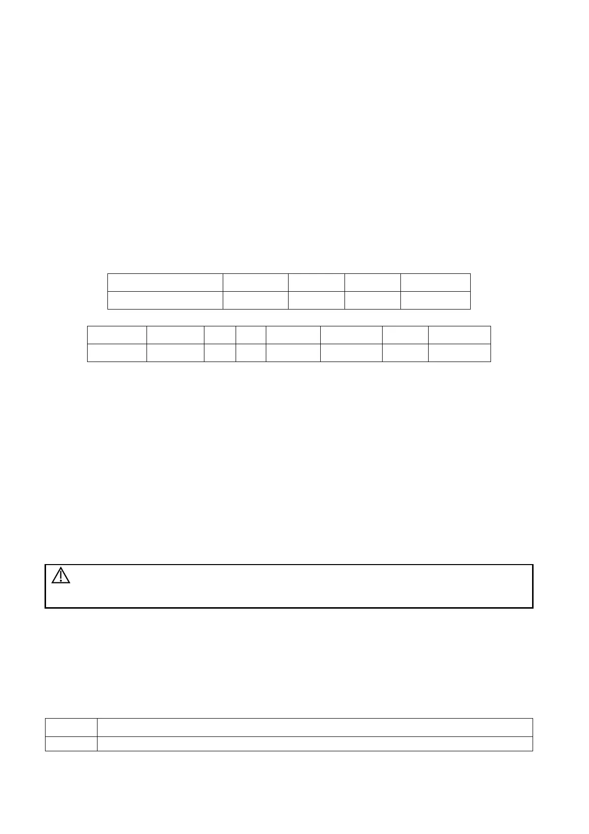
In TDI mode scanning, the image parameter area in the top-right corner of the screen displays the
real-time parameter values as follows:
TVI/TEI
Parameter
F G PRF WF
Meaning
Frequency Gain PRF Wall Filter
TVD
F G PRF WF SVD SV Angle
Frequency Gain PRF Wall Filter SV Position SV Size Angle value
5.9.3 TDI Image Optimization
In each TDI mode, the parameters that can be adjusted are similar to those in the color flow modes
(Color, PW, and Power). See the relevant sections for details. The following introduces the specific
items in TDI mode.
Tissue State
Description
This function is used for fast image optimization.
Operations
Adjust using the [Tissue State] item or mapping-menu item on the touch screen.
3 levels are provided: L, M, H.
5.9.4 TDI Quantitative Analysis (QA)
CAUTION:
TDI Quantitative Analysis results are provided for reference only, not for
confirming diagnoses. Compare the results with those of other machines,
or make diagnoses using non-ultrasound methods.
TDI QA is applied for TVI original data analysis, for evaluating the velocity change of the same cardiac
muscle with cardiac cycles.
The system provides 3 kinds of curves for quantitative analysis:
Speed – Time curve;
Strain – Time curve;
Strain Rate – Time curve.
Strain: Deformation and displacement of the tissue within the specified time.
Speed of the deformation, as myocardial variability will result in velocity gradient. Strain rate
 Loading...


