GE VOLUSON
i / VOLUSON
e
D
IRECTION KTI106052, REVISION 10 SERVICE MANUAL
Chapter 4 - Functional Checks 4-9
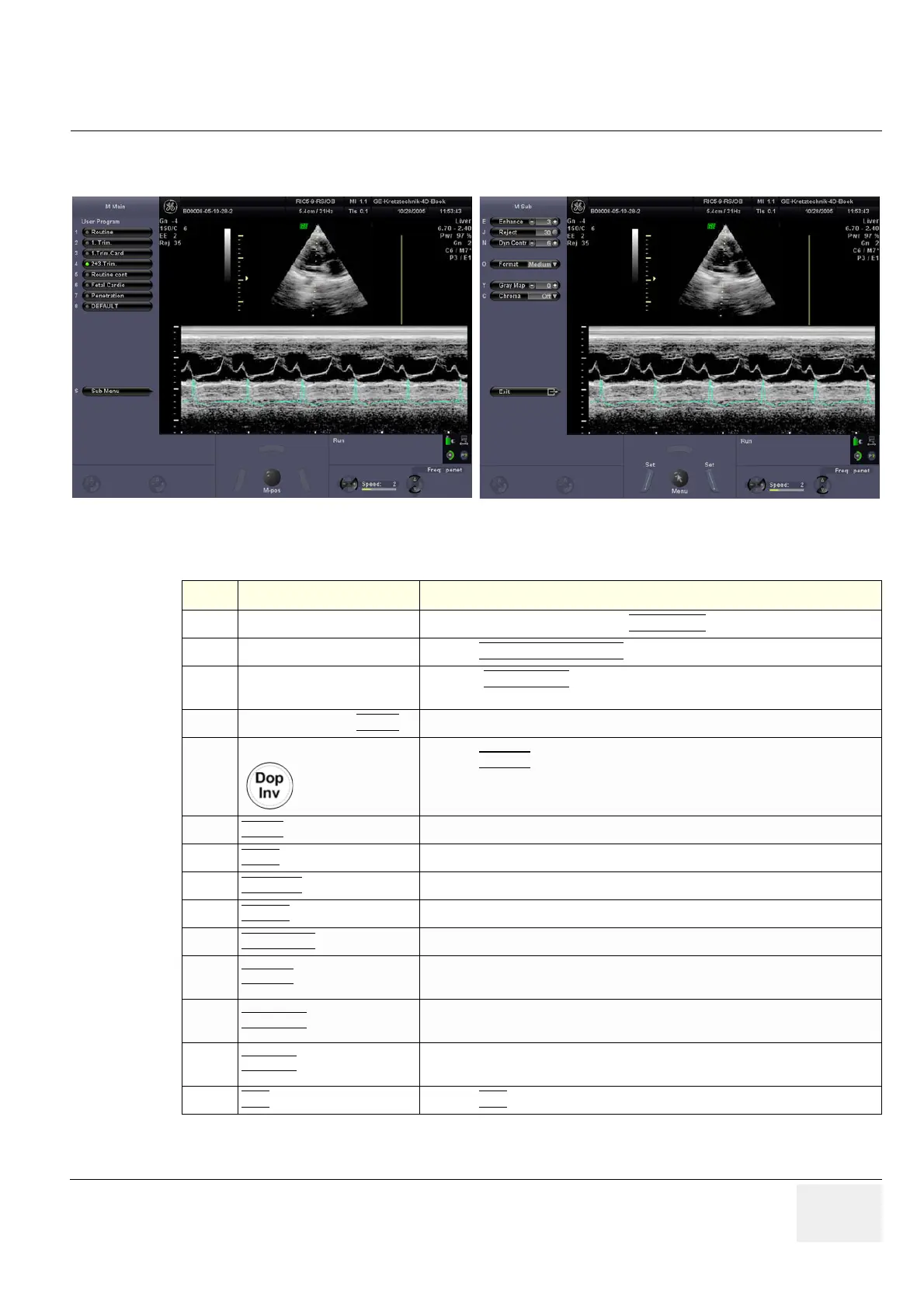
4-4-2 M Mode Checks
For further details refer to the Voluson i / Voluson e Basic User Manual, Chapter “M Mode”.
Figure 4-5 M Main and M Sub
Table 4-4 M Mode Functions
Step Task Expected Results
1
Cursor Position Adjust the M Cursor position with the TRACKBALL
in the 2D Single image.
2
Activation of M Mode Press the right or left trackball key
to activate both Modes (2D/M).
3
M Mode Gain
Rotate the ACTIVE MODE
rotary control to adjust the sensitivity (brightness) of the
entire M image.
4
M Mode Penetration DEPTH Directly Associated with 2D Mode Penetration Depth.
5
M Mode Trace Invert
Press the DOP INV key on the control panel to invert the M mode trace from up to
down in the M mode display area.
(The Invert function is only available with endovaginal probes.)
6
SPEED Press the Soft-menu buttons to select different sweep speeds.
7
FREQ:
Common with 2D Mode Freq: Resol. Harm.Freq. in case of Harm. Imaging.
8
ENHANCE
Due to this function a finer, sharper impression of the image is produced.
9
REJECT
It determines the amplitude-level below which echoes are not displayed (rejected).
10
DYN CONTR Dynamic enhances a part of the grayscale to make it easier to display pathology.
11
FORMAT
For selection of three different changes the display of the ratio between the
grayscale image and M-Mode spectrum.
12
GRAY MAP
A gray map determines the displayed Brightness of an echo in relationship to its
amplitude.
13
CHROMA
This defines the relation between echo amplitude (input) and Chroma value (color
tone and saturation) in a look-up table.
14
EXIT
Press the EXIT key on the control panel to exit the M Sub menu.

 Loading...